Coral larvae (Acropora cytherea) settled on aragonite plug.
Photo: Eneour Puill-Stephan
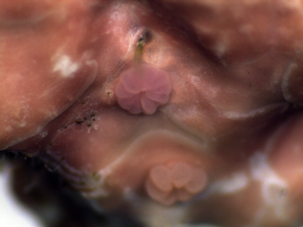
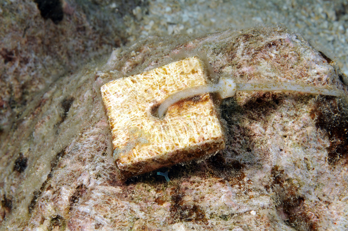
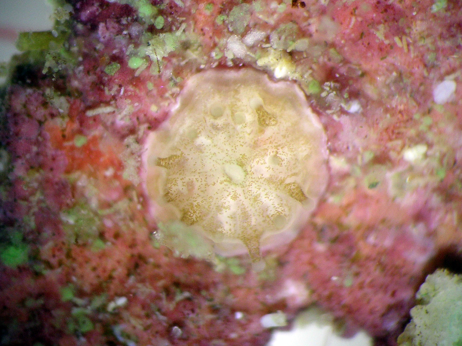
Coral settler (Favia fragrum) on a preconditioned limestone settlement tile.
Photo: Maggy Nugues
Coral larvae searching for a settlement substrate in a laboratory container.
Photo: Maggy Nugues
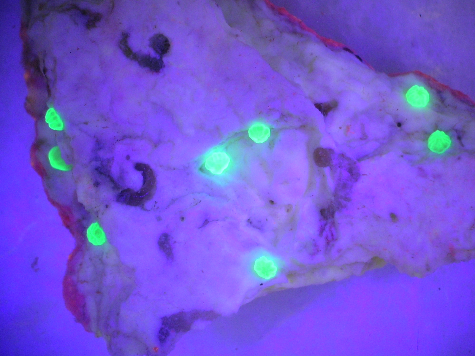
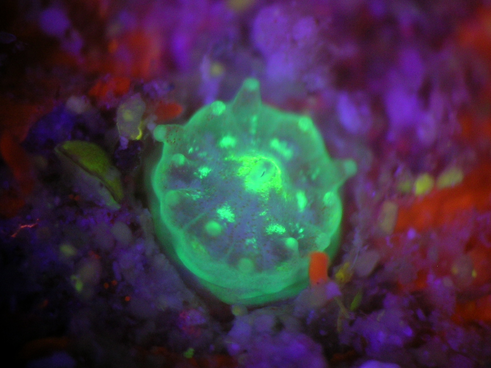
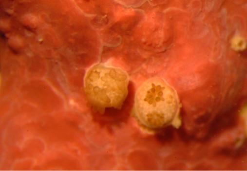
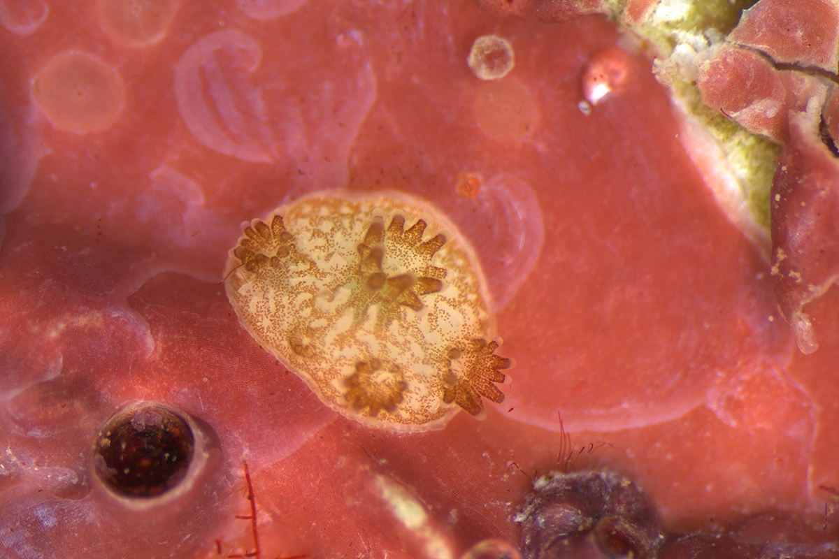
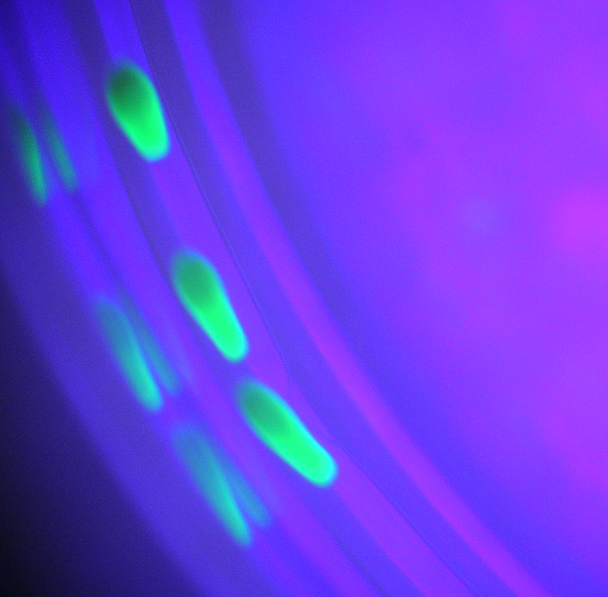
Coral settlers (Favia fragrum) on small chip of crustose coralline algae photographed under blue UV light. UV light helps with detecting and counting tiny settlers.
Photo: Maggy Nugues

Adult coral (Orbicella faveolata) emitting gamete bundles during mass spawning event in Puerto Rico. Gamete bundles can be collected and raised in the lab to larval stages for coral restoration.
Photo: Maggy Nugues
Coral settlers on the crustose coralline alga Titanoderma prototypum, a well known settlement-inducer.
Photo: Robert Steneck

Crustose coralline alga photographed in situ. These algae are notoriously difficult to identify.
Photo: Maggy Nugues
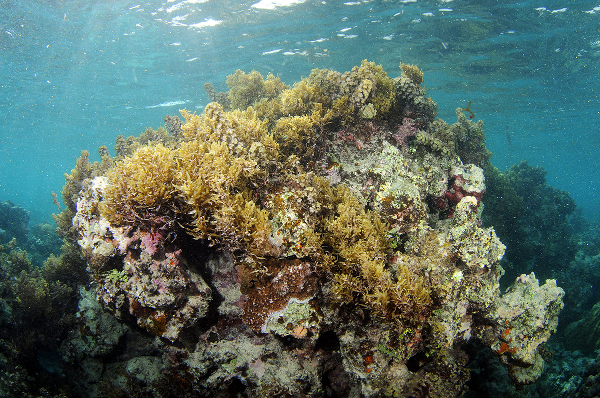
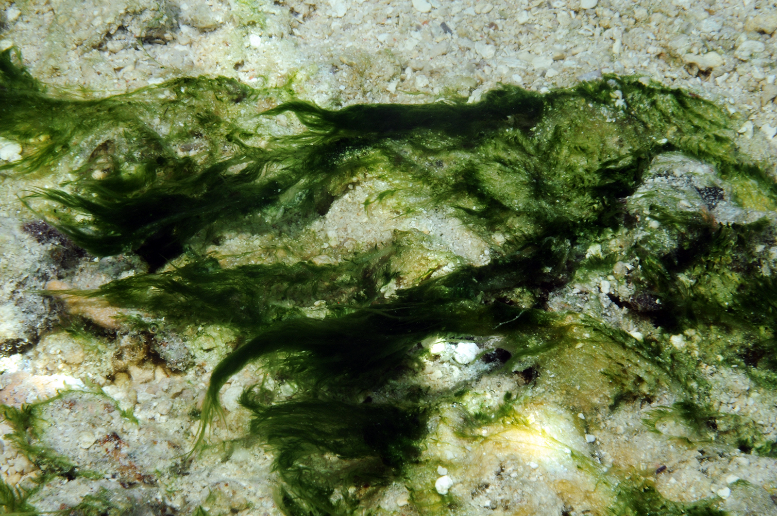
Coral bommy covered with macroalgae in the lagoon of Moorea.
Photo: Maggy Nugues
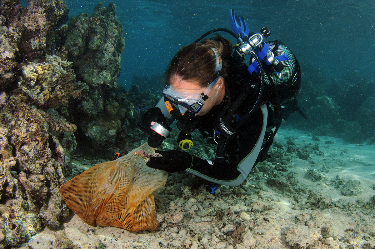
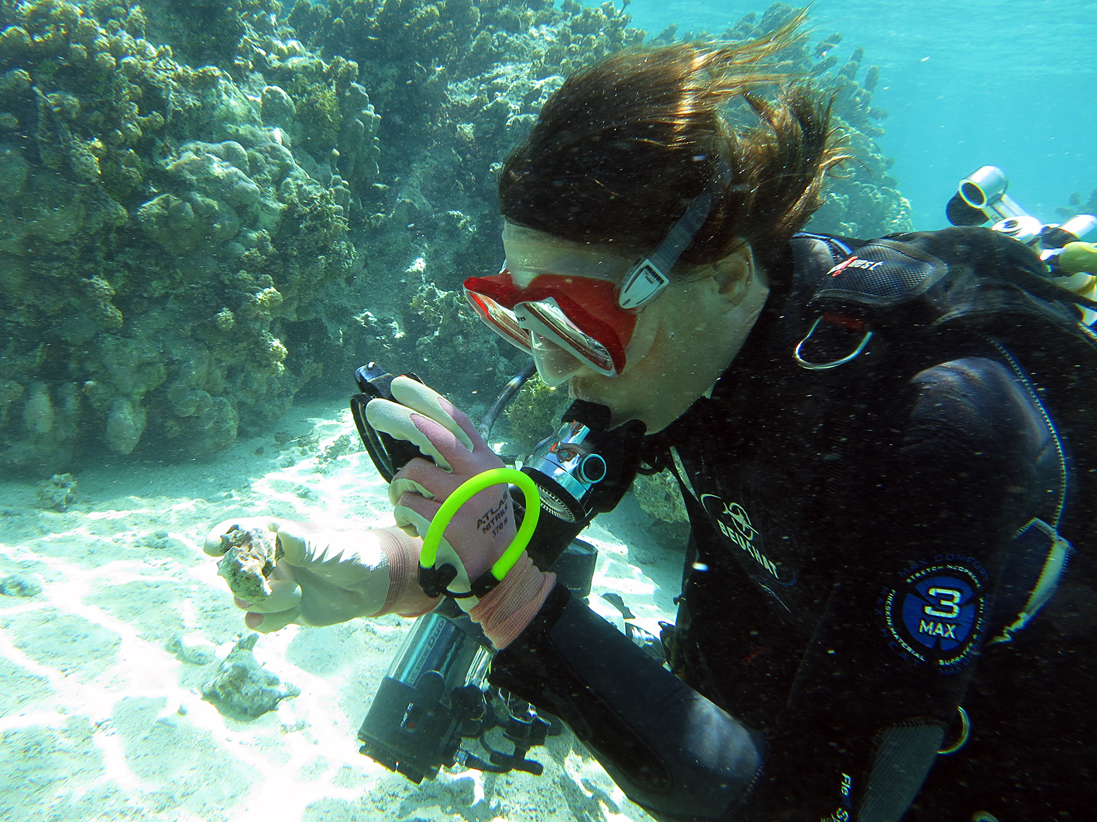
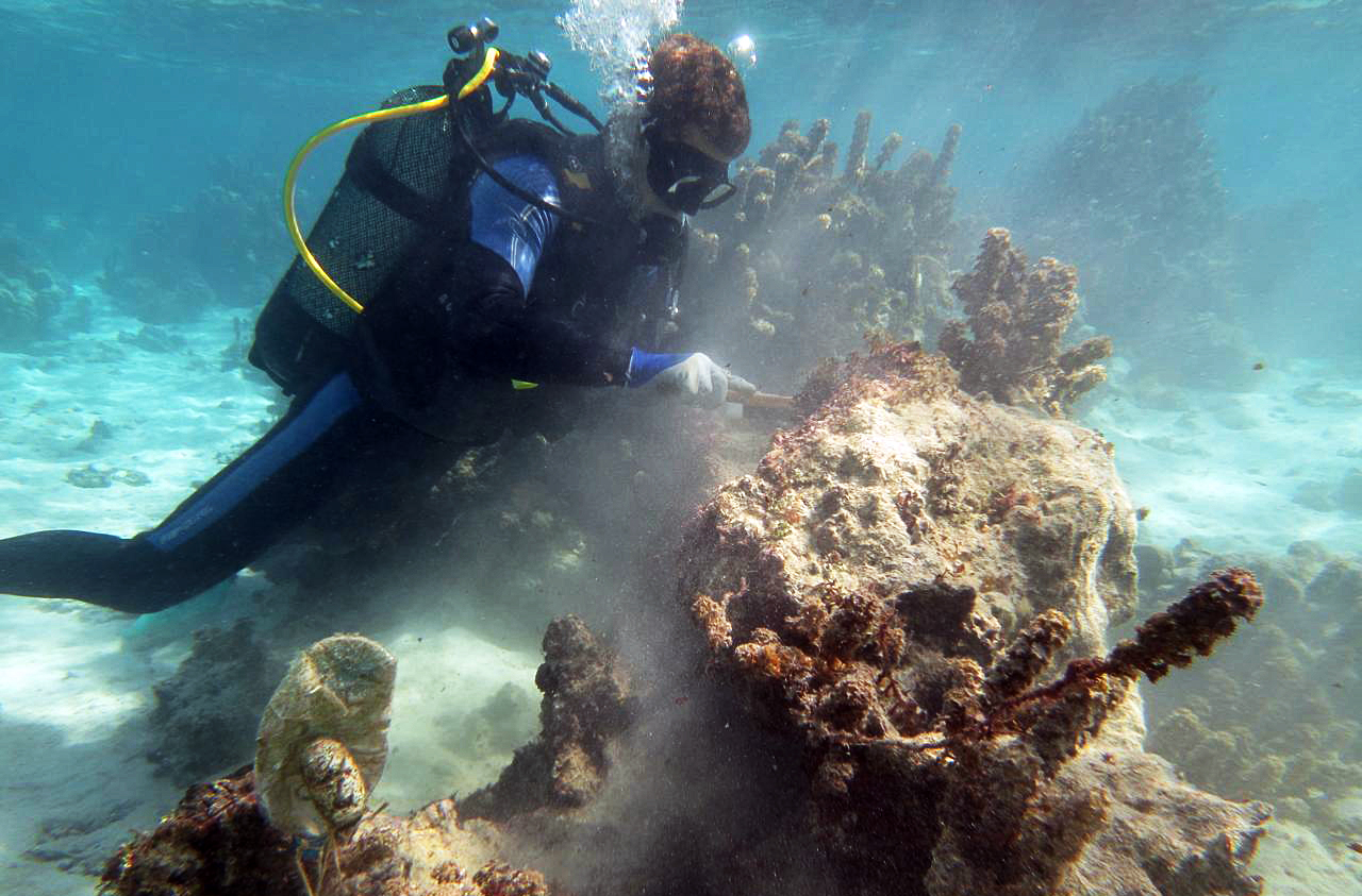
Hendrikje collecting crustose coralline algae.
Photo: Maggy Nugues

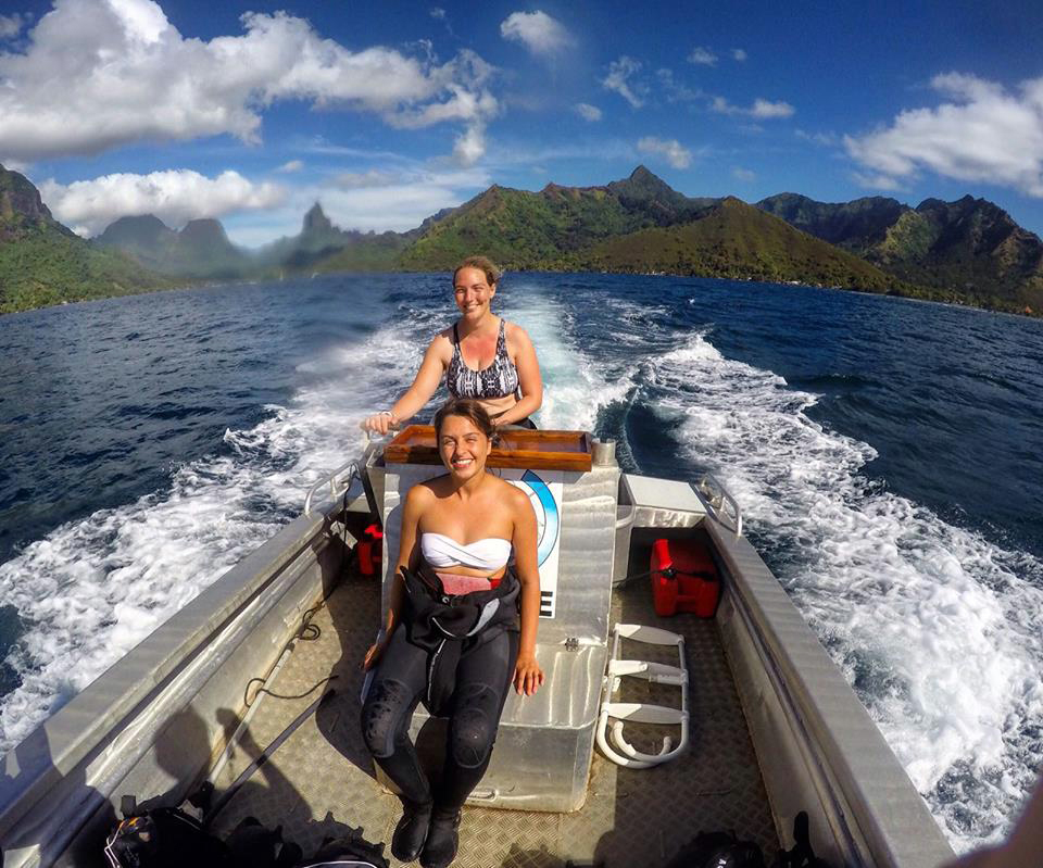
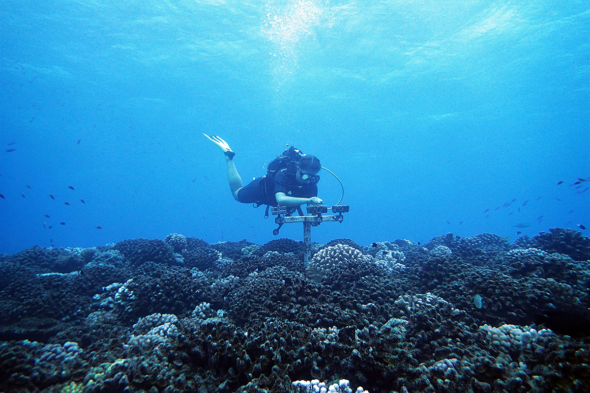
Getting out in the field from research station.
Photo: Maggy Nugues
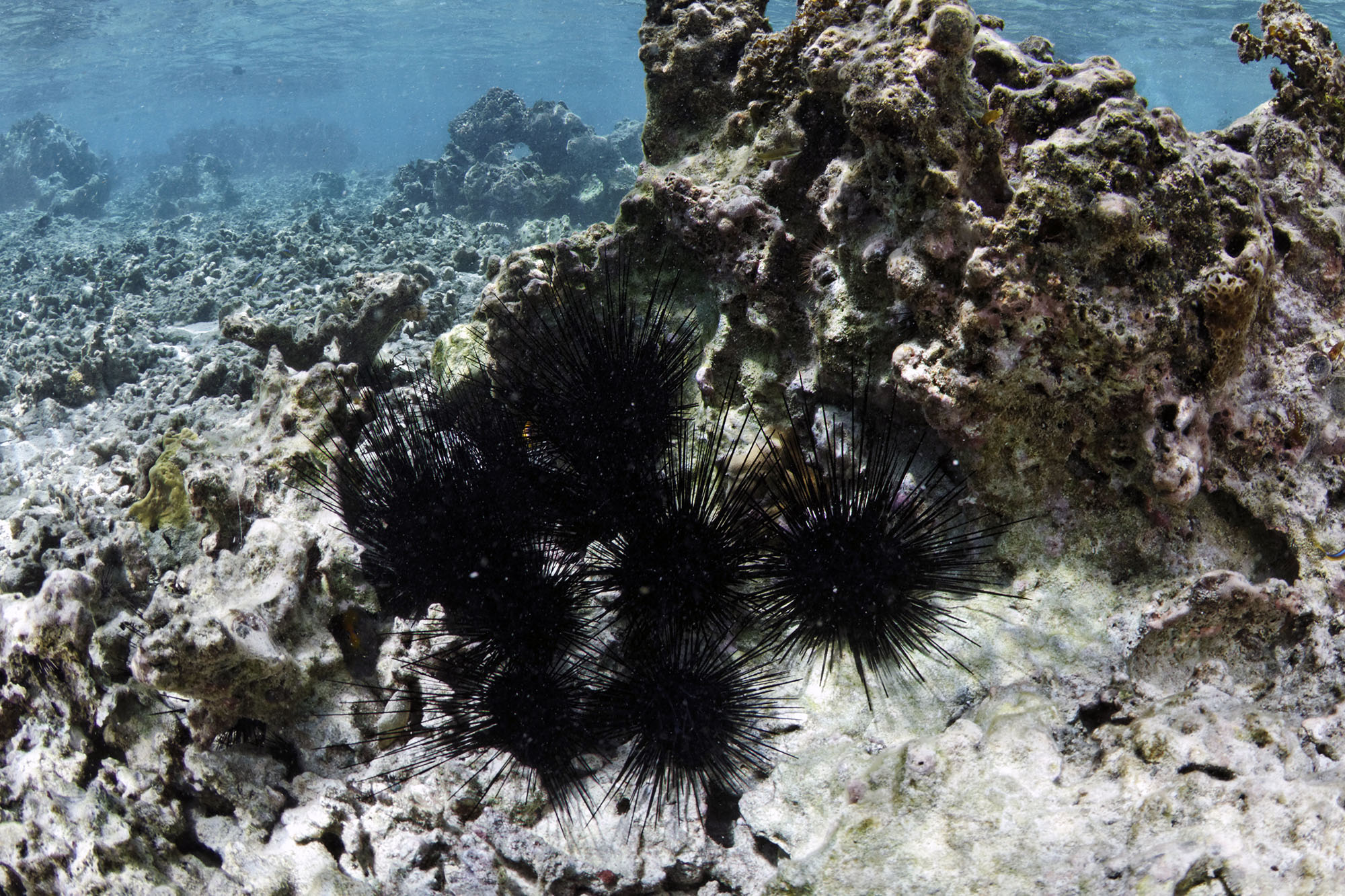
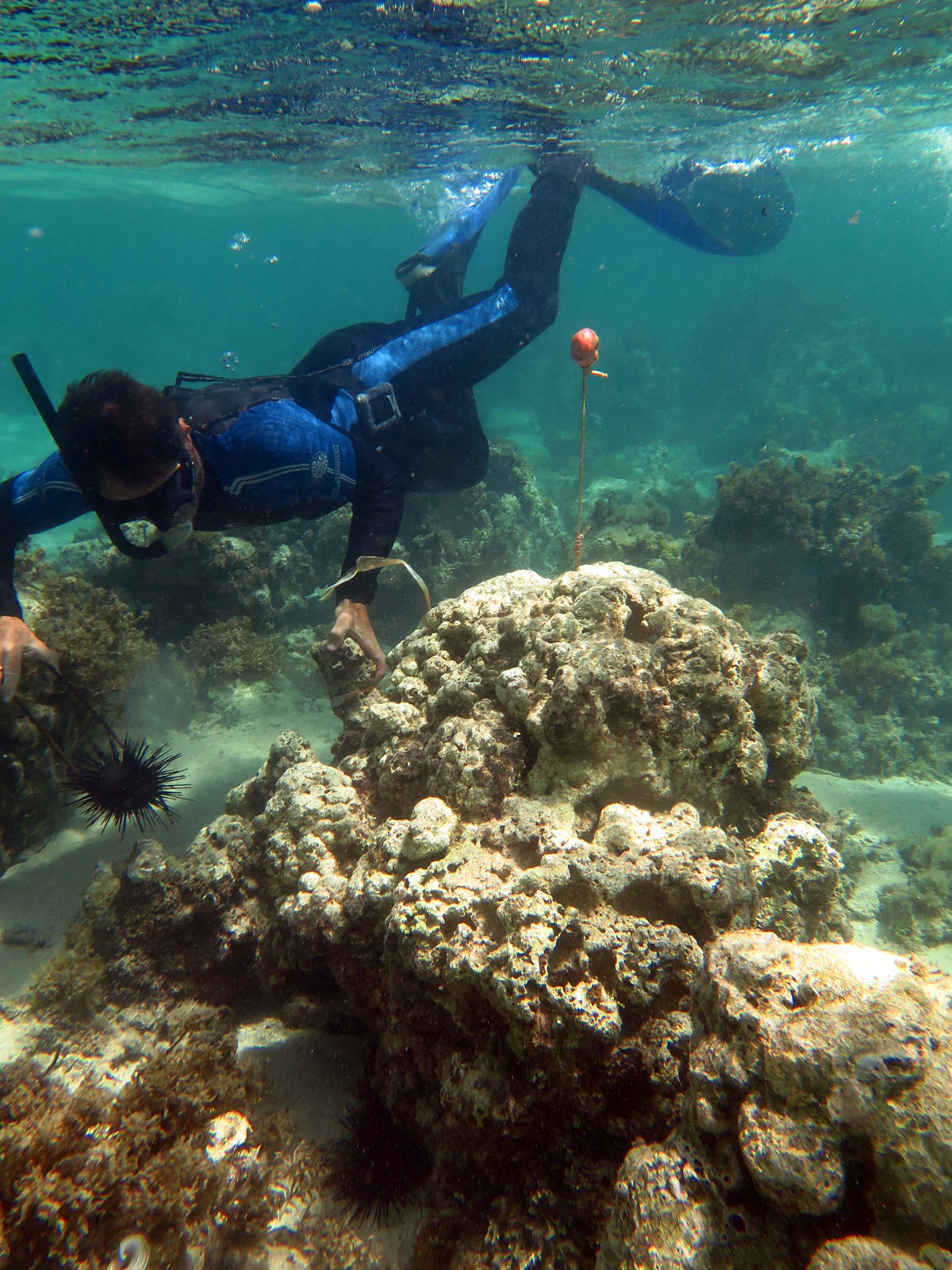
Herbivorous sea urchins in the lagon of Moorea. Sea urchins can act as facilitators of coral recruitment.
Photo: Gilles Siu
Fabio Bulleri deploying Diadema in the field.
Photo: Julien Gasc

Natural stands of Turbinaria ornata in the field.
Photo: Fabio Bulleri
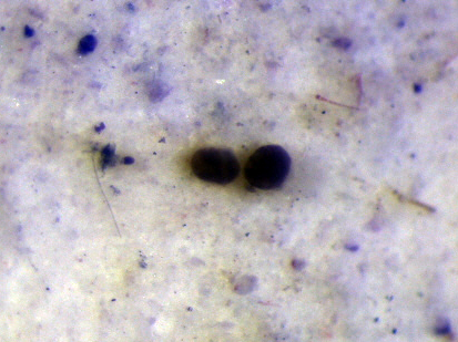
Two settled germlings of Turbinaria ornata in the lab.
Photo: Fabio Bulleri

Chloé transplanting a Pocillopora damicornis colony.
Photo: Hugo Bischoff
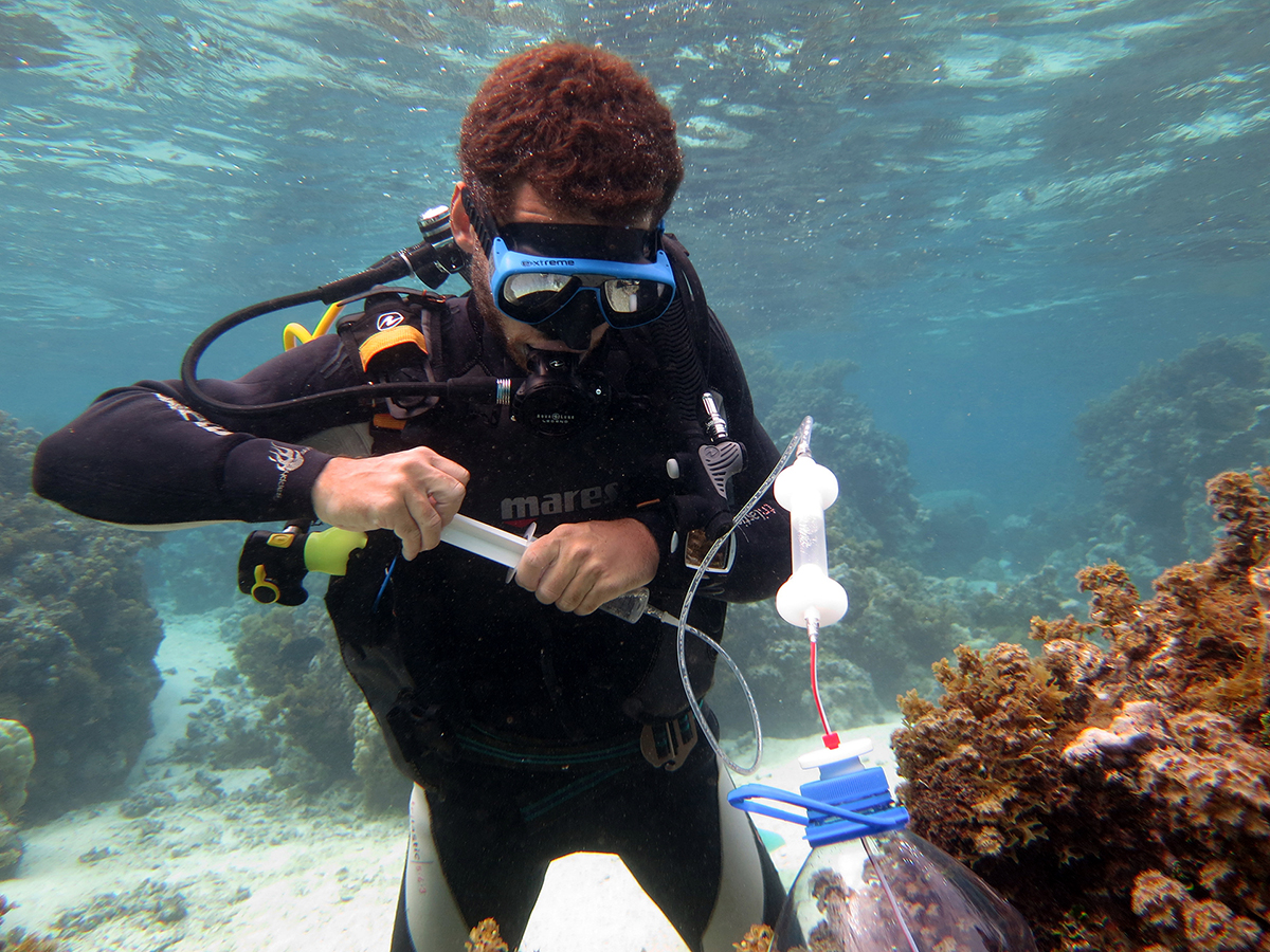
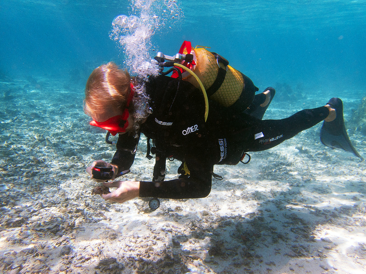
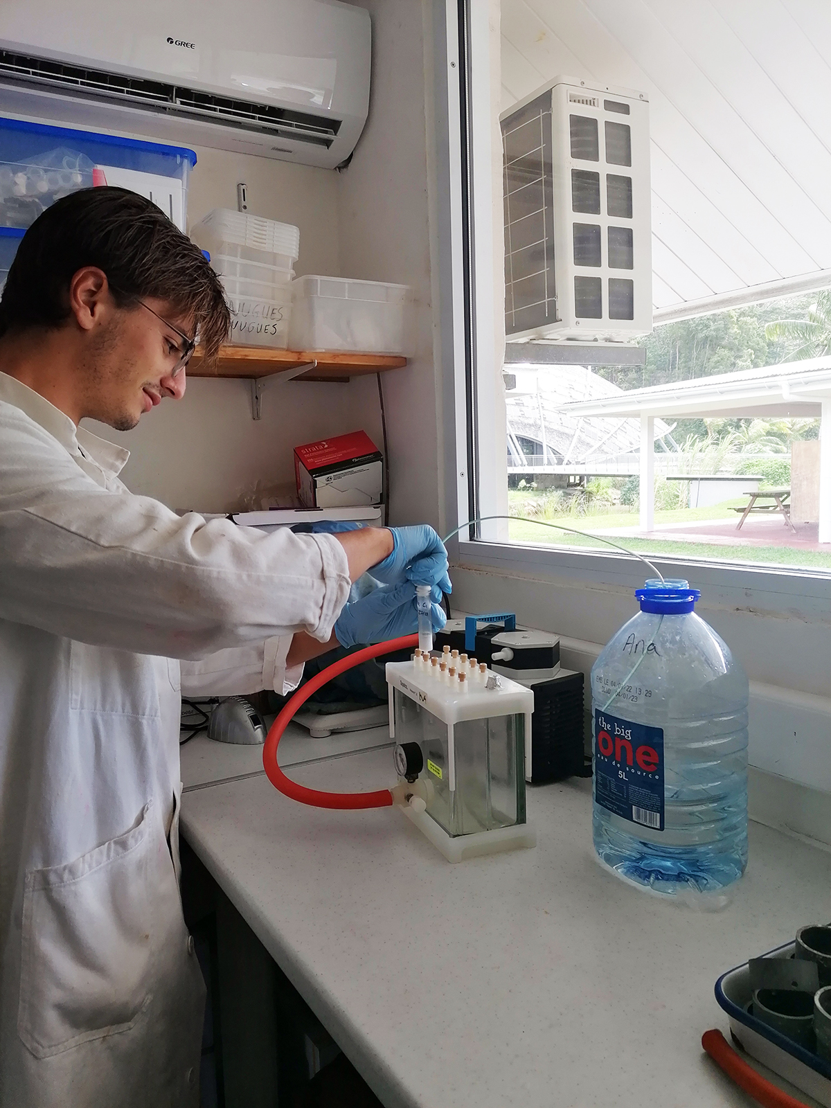
Hugo pulling a syringe for in-situ extraction of chemical compounds from the water column.
Photo: Chloé Pozas-Schacre
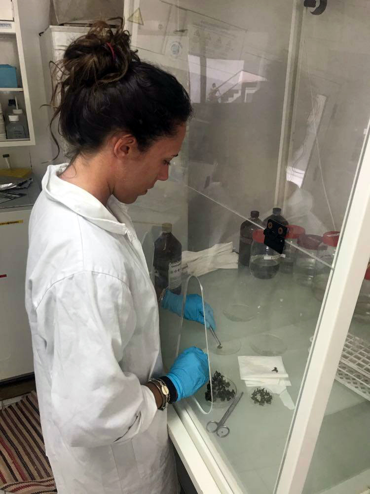
Chloé dipping fragments of Turbinaria ornata in methanol to extract surface metabolites.
Photo: Hugo Bischoff
Hendrikje and Chérine Baumgartner going out in the field.
Photo: Mischa Streekstra
Hendrikje collecting coral larvae.
Photo: Sonora Meiling
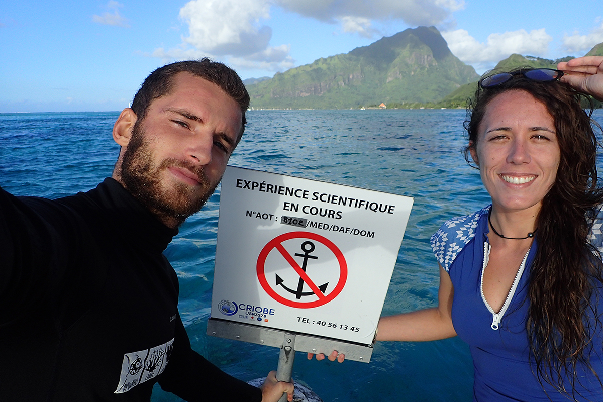
Chloé & Hugo in front of their “no anchorage” sign.
Photo: Hugo Bischoff
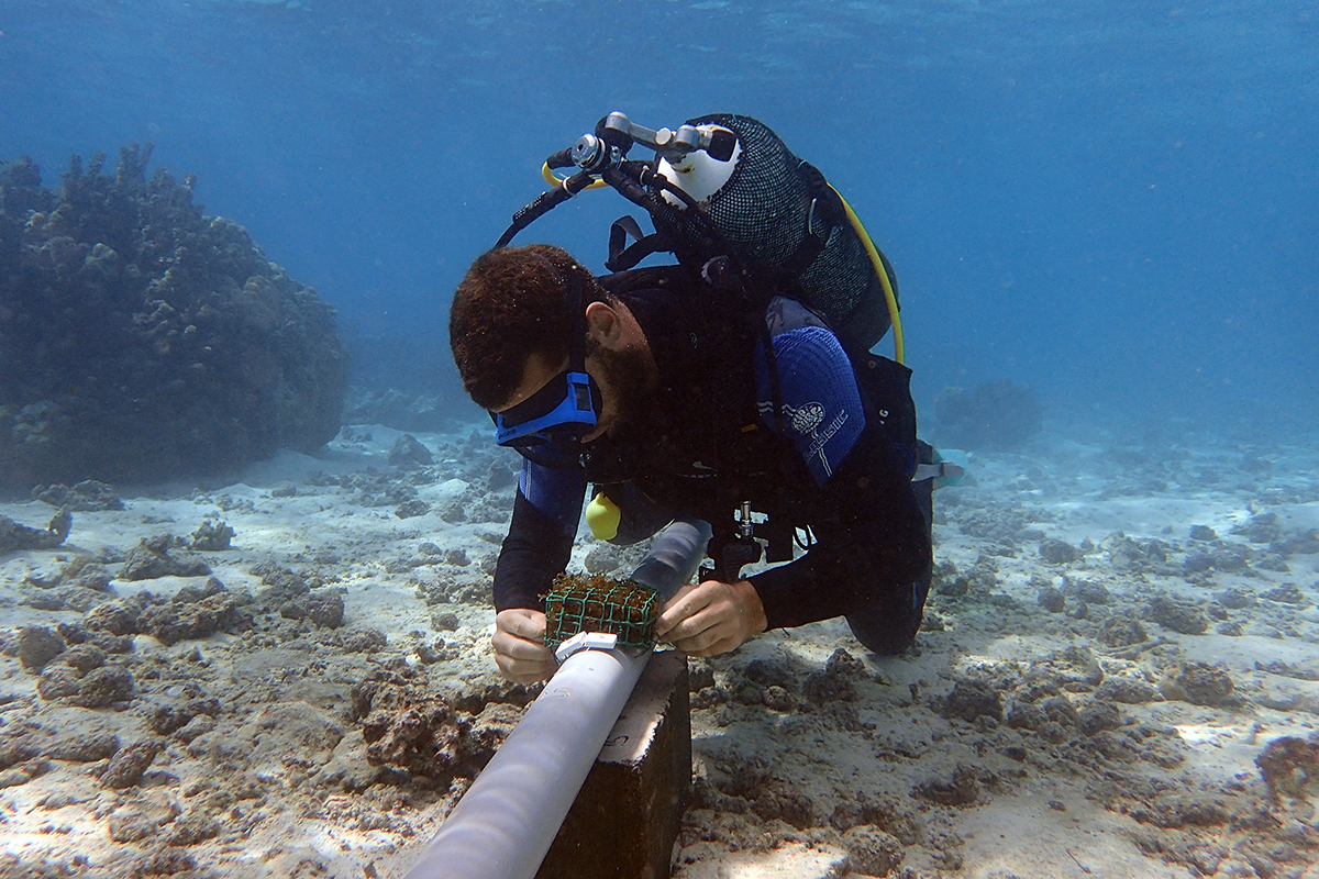
Hugo installing macroalgae on one of the structures of the second experiment.
Photo: Chloé Pozas-Schacre

Chloé removing macroalgae from an experimental bommie.
Photo: Hugo Bischoff

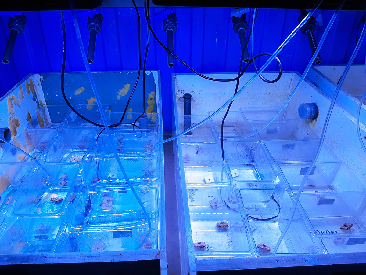
Experimental room with the glass cylinders.
Photo: Chloé Pozas-Schacre
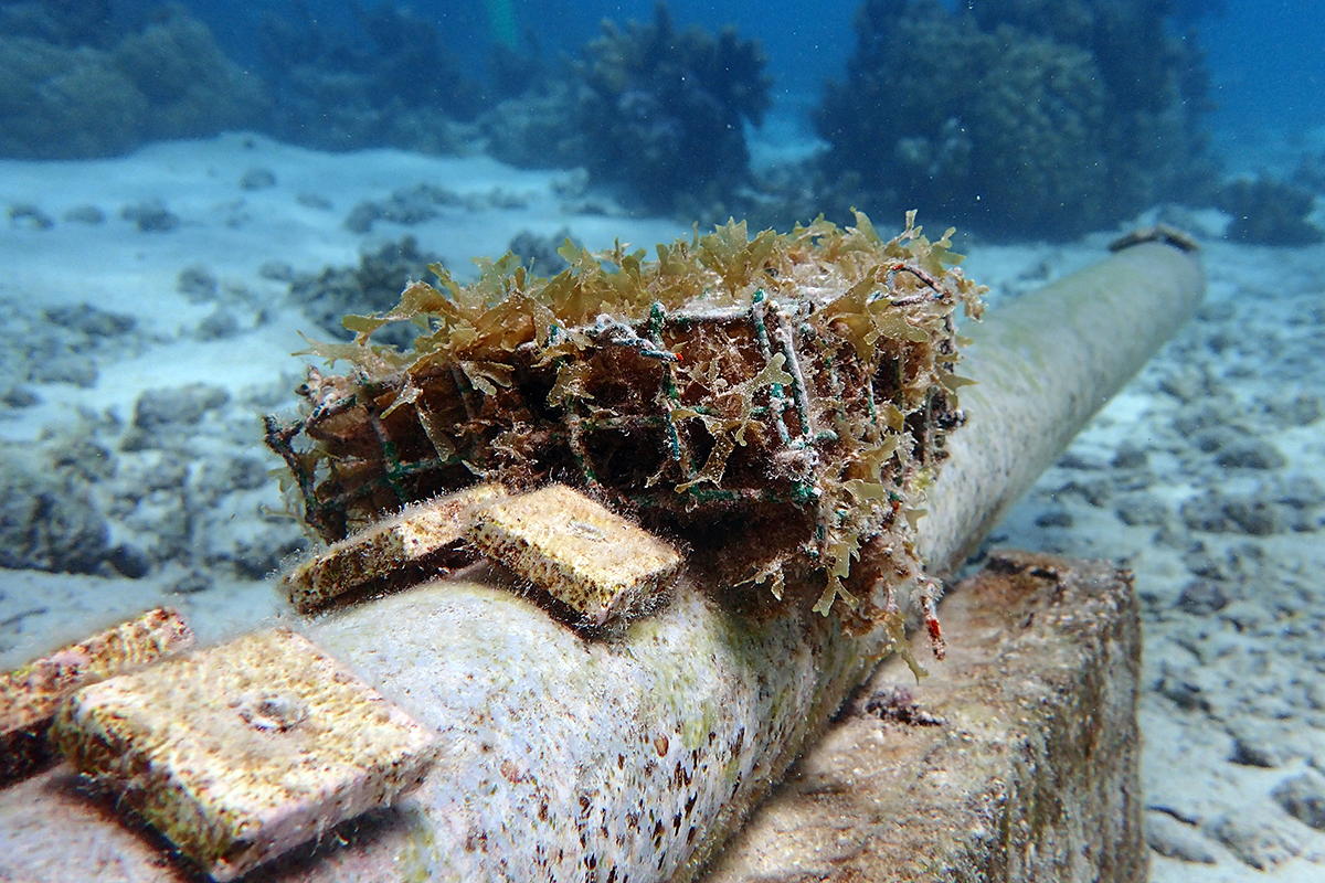
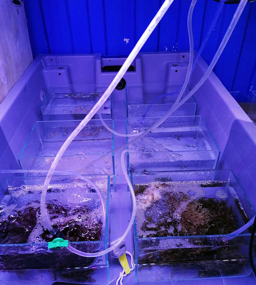
Structure holding tiles conditioned in the presence of Dictyota bartayresiana.
Photo: Chloé Pozas-Schacre
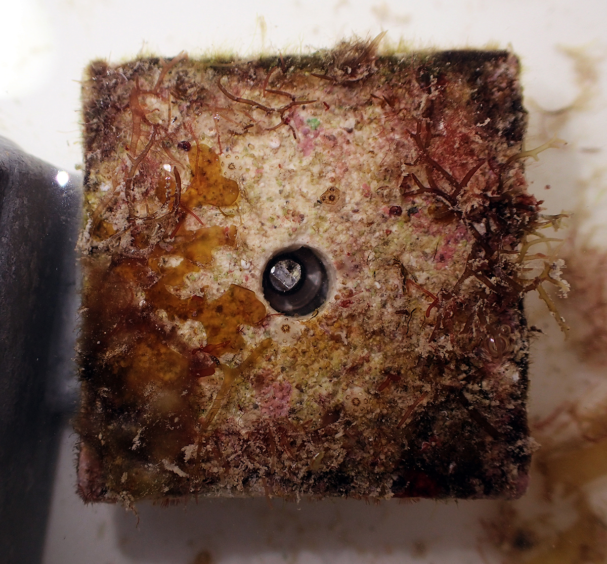
Cryptic side of a recruitment tile. Can you find the coral recruits? Click on the photo and zoom in.
Photo: Chloé Pozas-Schacre

Natural pocilloporid recruits interacting with crustose coralline algae.
Photo: Maggy Nugues

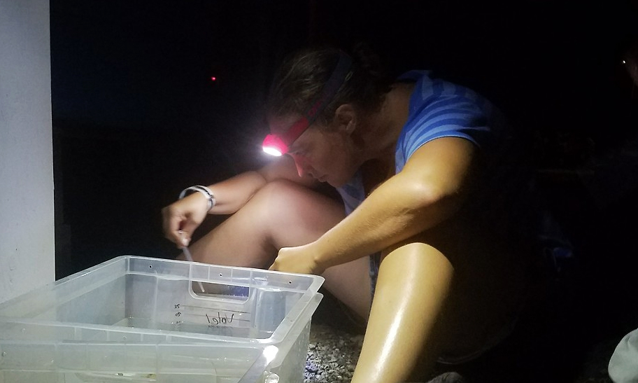
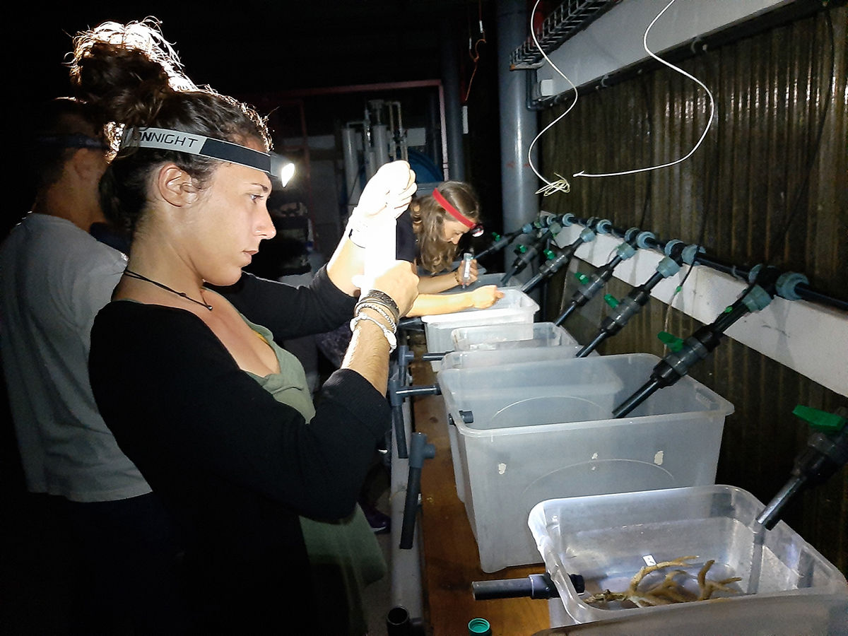
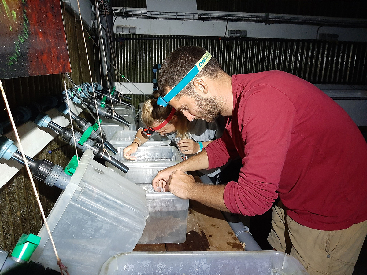
Hendrikje and Yann Lacube checking up experimental dishes.
Photo: Isabel Ender
Experimental petridishes showing coral recruits in contact with crustose coralline algae.
Photo: Maggy Nugues
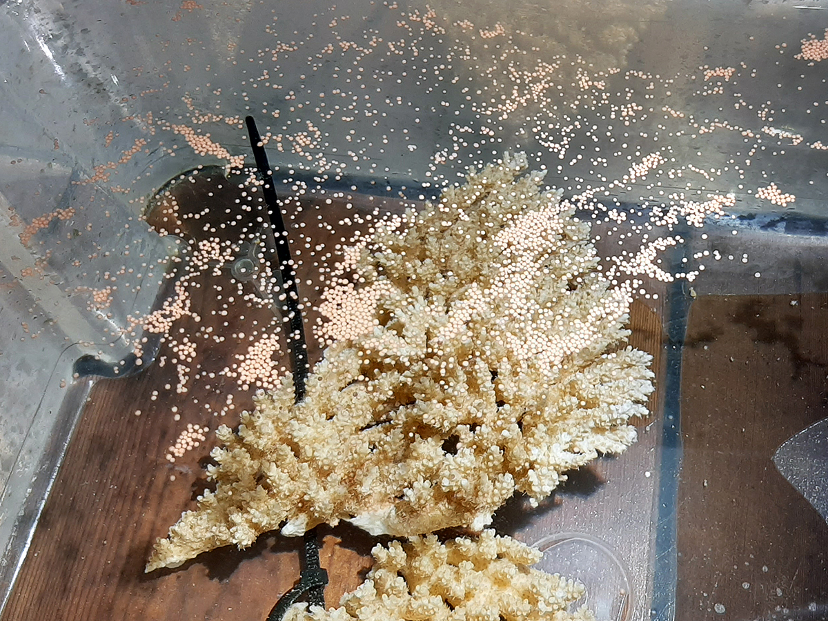
Acropora cytherea colony releasing egg-sperm bundles during spawning.
Photo: Maggy Nugues
Chloé concentrating egg-sperm bundles inside a collecting tube during spawning night.
Photo: Maggy Nugues

Camille running larval behavioral choice experiment.
Photo: Maggy Nugues
Two Acropora recruits settled on Titanoderma chip in the high oxygen end of the hypoxitron.
Photo: Hendrikje Jorissen
Pierre-Louis searching for the crustose coralline algae Titanoderma.
Photo: Camille Vizon
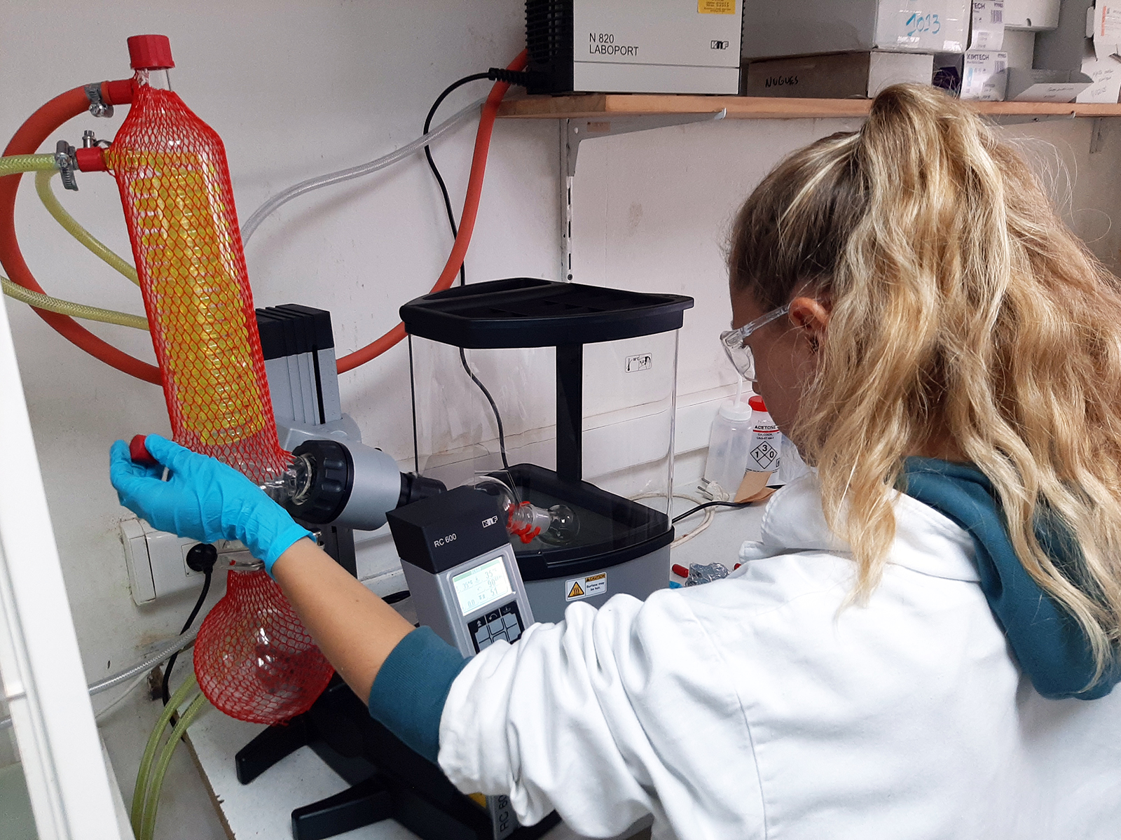
Preparation of algal exudates in the laboratory.
Photo: Camille Vizon

Preparation of algal exudates in the laboratory.
Photo: Camille Vizon

Lettuce like crustose coralline algae.
Photo: Maggy Nugues

Hugo placing CCA fragments in silica gel for genetic characterisation.
Photo: Maggy Nugues
Hugo placing CCA fragments in silica gel for genetic characterisation.
Photo: Maggy Nugues
Camille and Hugo collecting gamete bundles.
Photo: Maggy Nugues
Conditioning of experimental tile used in coral settlement experiments.
Photo: Maggy Nugues
Conditioning of experimental tile used in coral settlement experiments.
Photo: Maggy Nugues

Open Day with crustose coralline algae stand.
Photo: Annaïg Le Guen
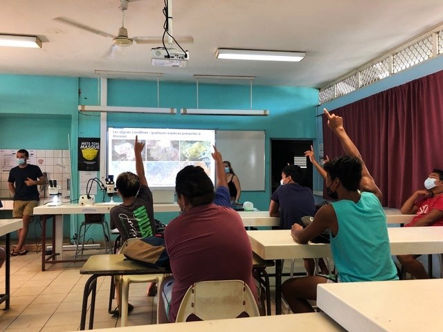
Camille and Hugo giving a lecture to middle school pupils.
Photo: Vie Stabile
Camille and Hugo giving a lecture to middle school pupils.
Photo: Vie Stabile

Camille adding chemical extracts to dead algal fragments.
Photo: Camille Vizon
Camille evaporating chemical extracts.
Photo: Maggy Nugues
One-day old coral spat on the crustose coralline algae Titanoderma prototypum.
Photo: Camille Vizon
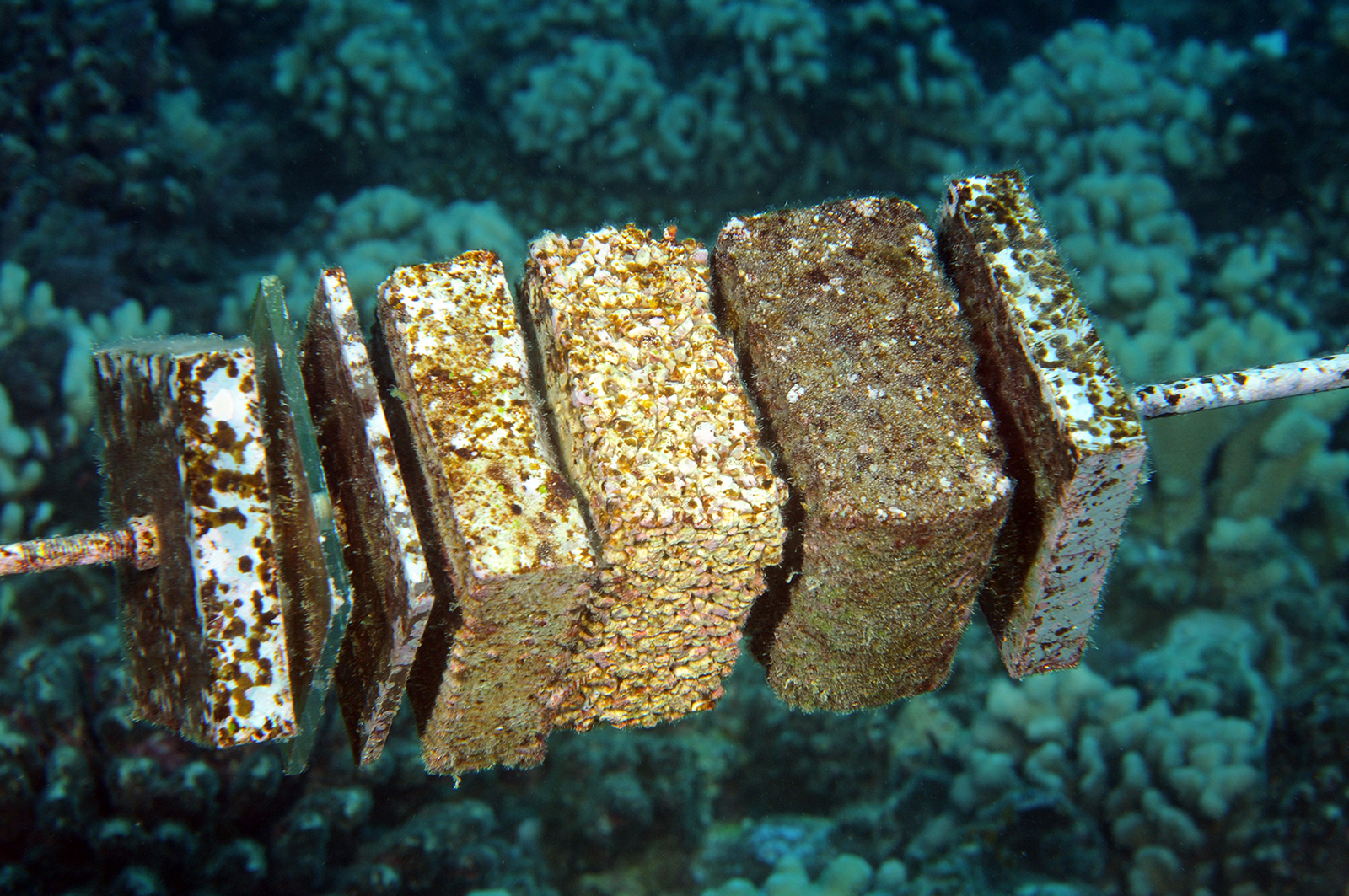
Stacks of vertically oriented tiles made of different materials.
Photo: Maggy Nugues
Camille Leonard replacing recruitment tiles.
Photo: Hugo Bischoff
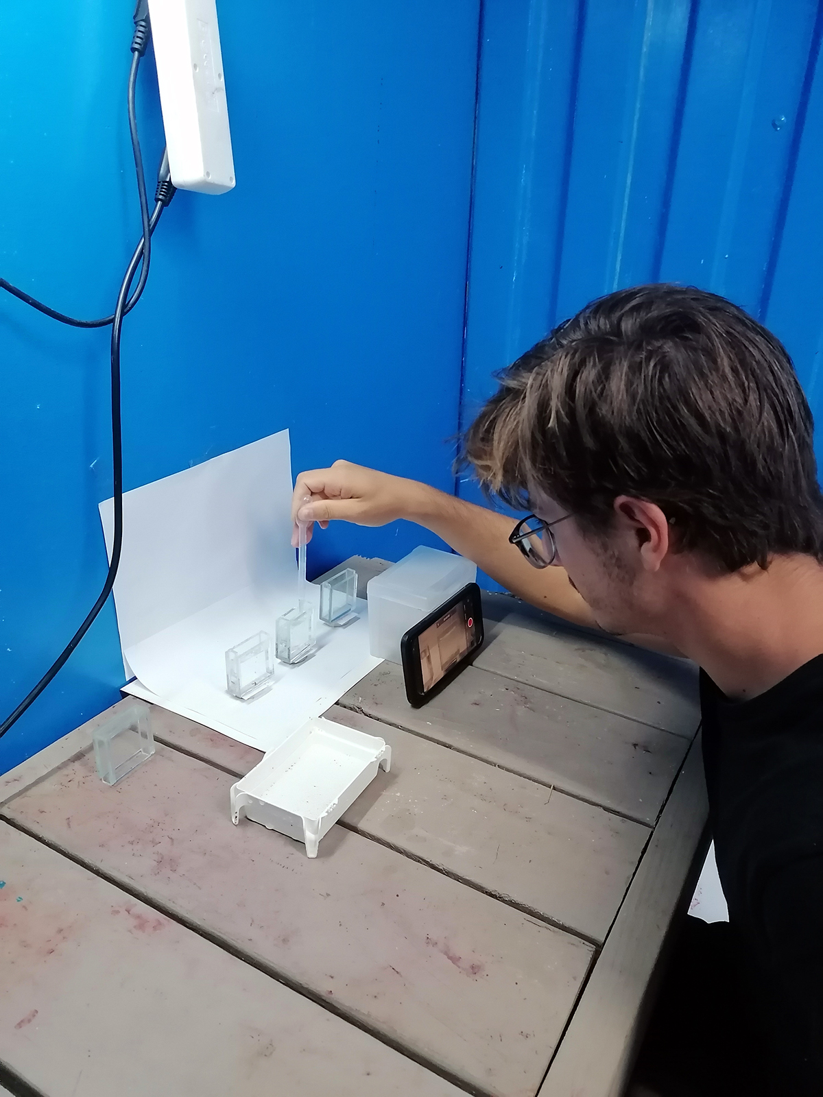
Larval behavioral assays using video tracking.
Photo: Camille Vizon
Larval behavioral assays using video tracking.
Photo: Camille Vizon
Vacuum filtration of algal exudates.
Photo: Camille Vizon
Benthic Anabaena sp. mat.
Photo: Maggy Nugues
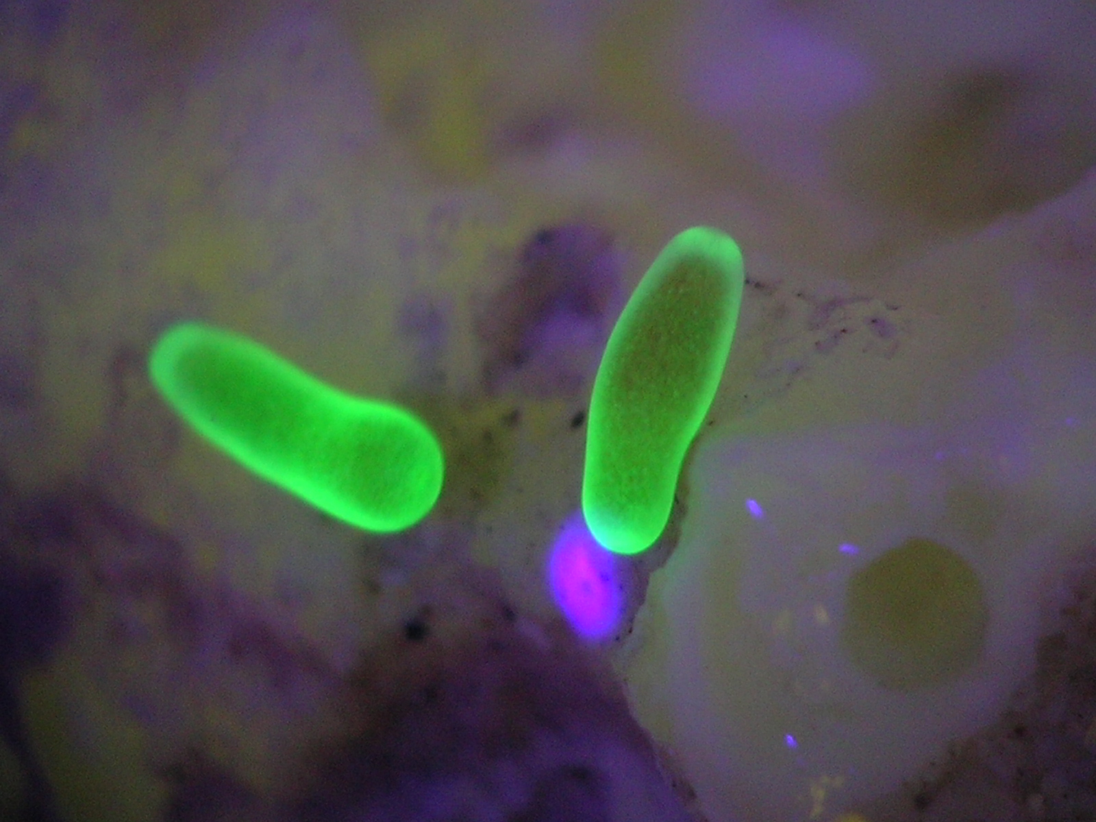
Coral larvae exploring the experimental dish photographed under blue UV light.
Photo: Maggy Nugues
Young coral recruits.
Photo: Maggy Nugues

Hugo Bischoff adjusting Diadema densities on the experimental bommies.
Photo: Maggy Nugues
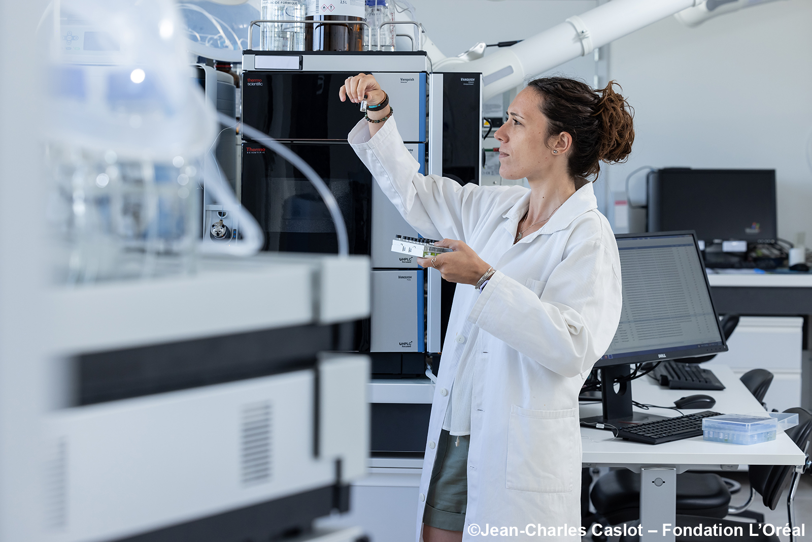
Chloé in the chemistry lab.
Photo: Jean-Charles Caslot
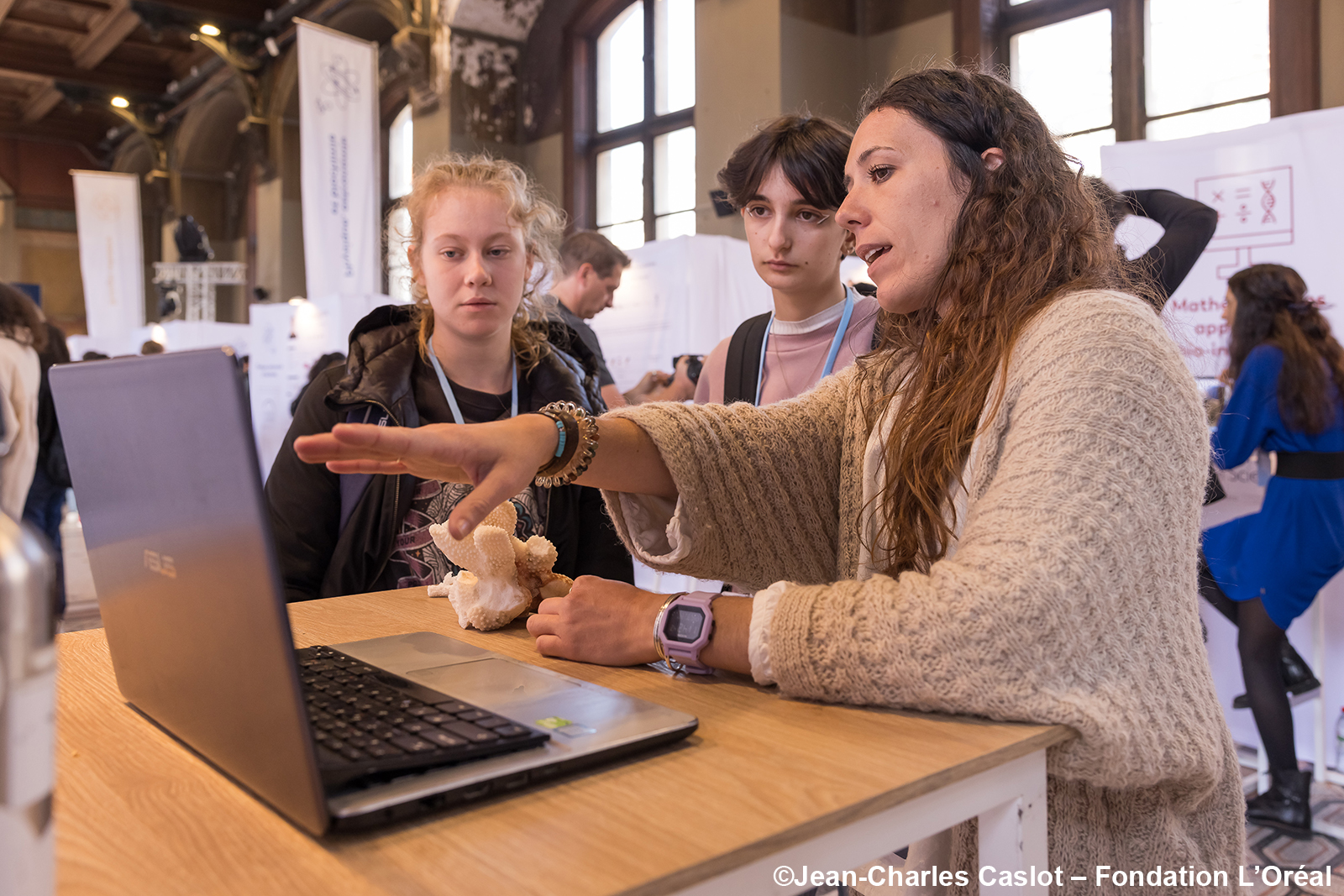
Chloé explaining her work to school pupils.
Photo: Jean-Charles Caslot
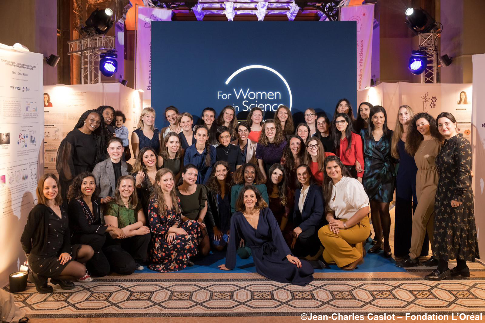
Fondation L’Oreal-UNESCO French Young Talents 2022.
Photo: Jean-Charles Caslot
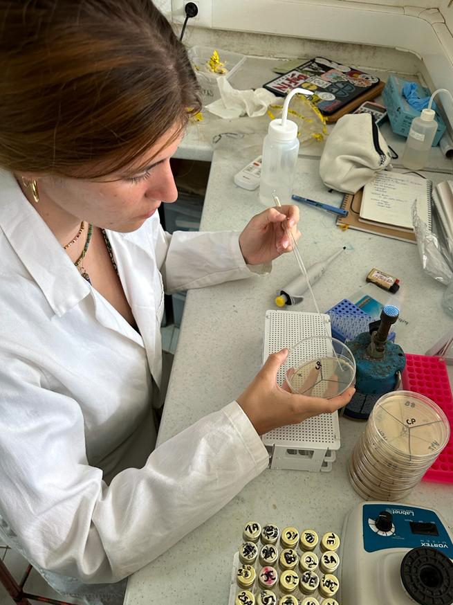
Cassandra streaking for isolation on an agar plate.
Photo: Clément Esclavy
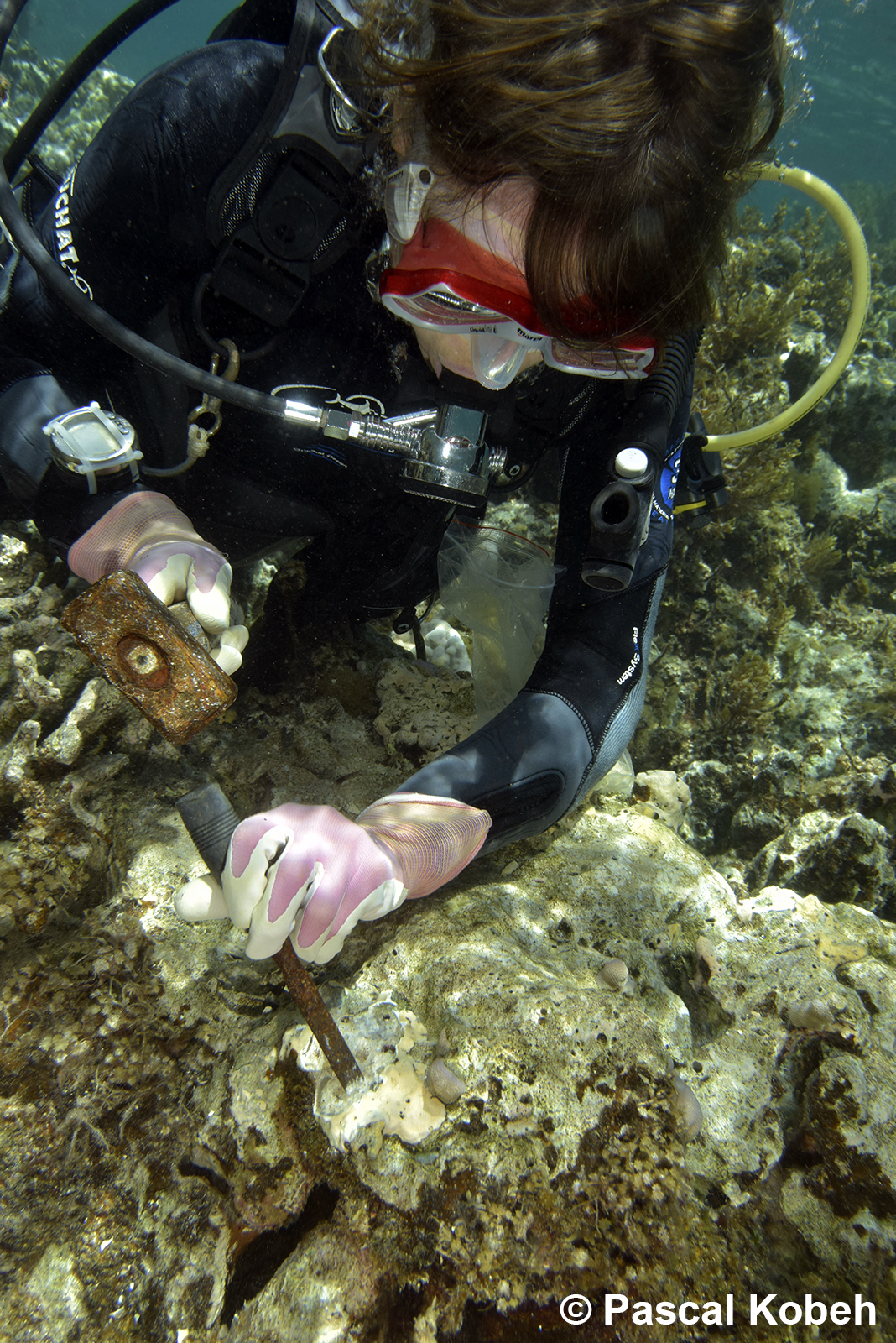
Maggy sampling crustose coralline algae.
Photo: Pascal Kobeh
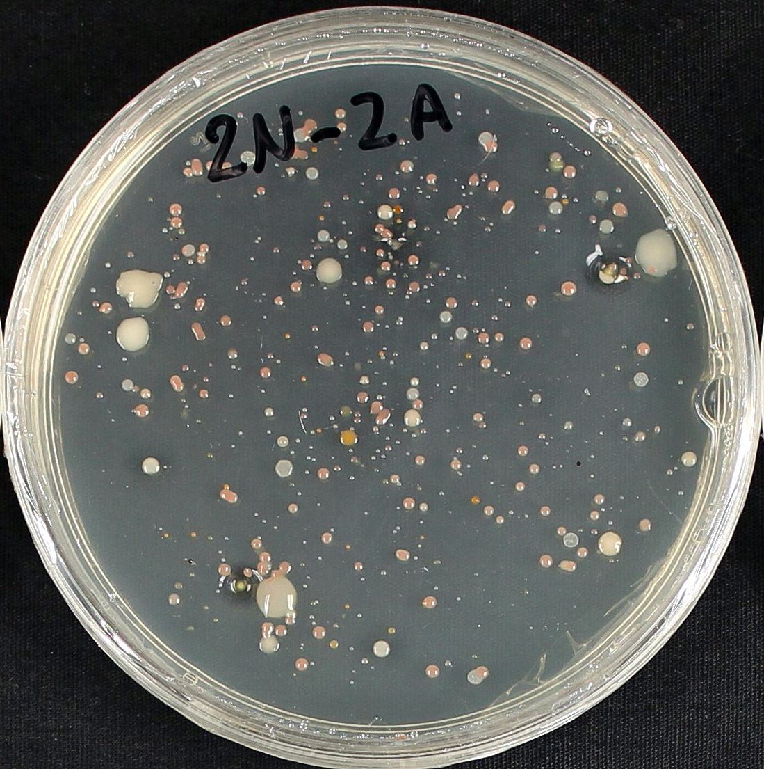
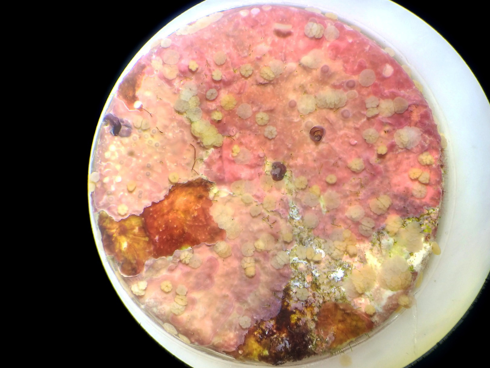
Culture plate after 5 days.
Photo: Cassandra Le Bélicard
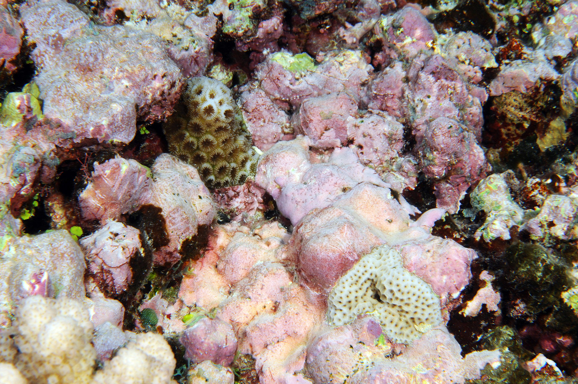
Crustose coralline algal dominated reef substratum in Moorea.
Photo: Maggy Nugues
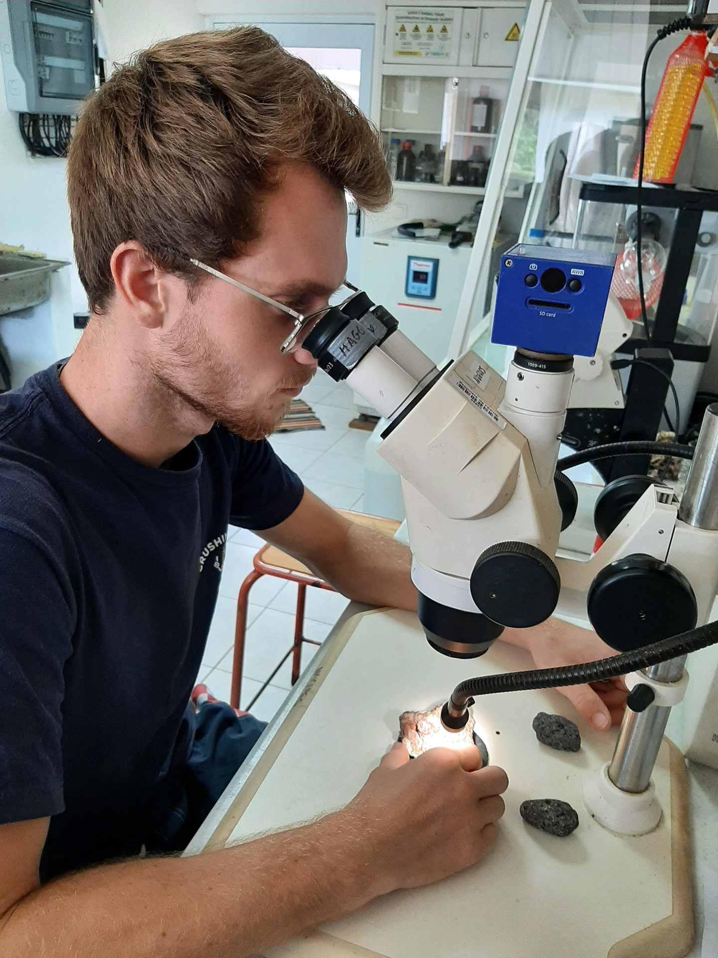
Clément observing a coral recruit in the lab.
Photo: Maggy Nugues
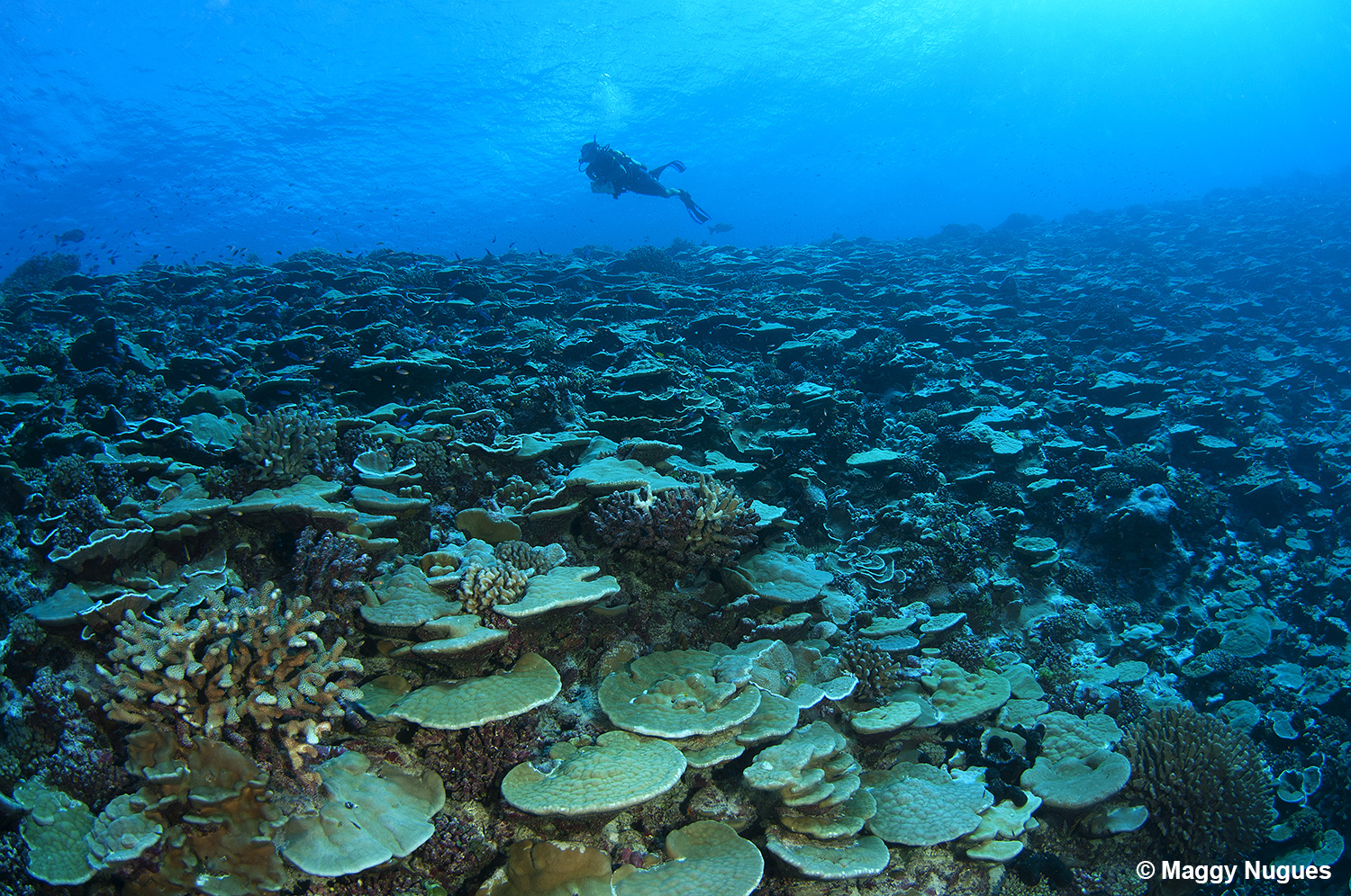
Coral reef of Rangiroa atoll, French Polynesia.
Photo: Maggy Nugues
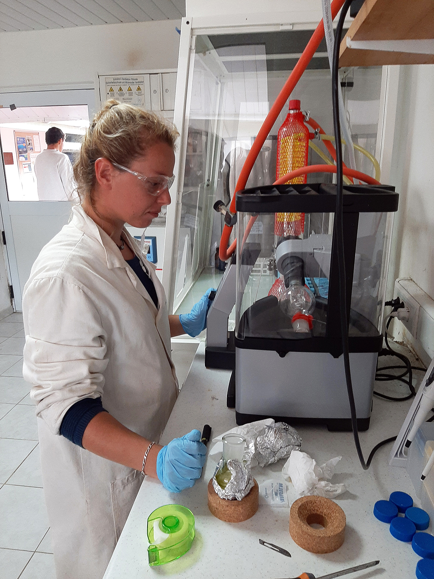
Evaporation of extraction solvent.
Photo: Maggy Nugues
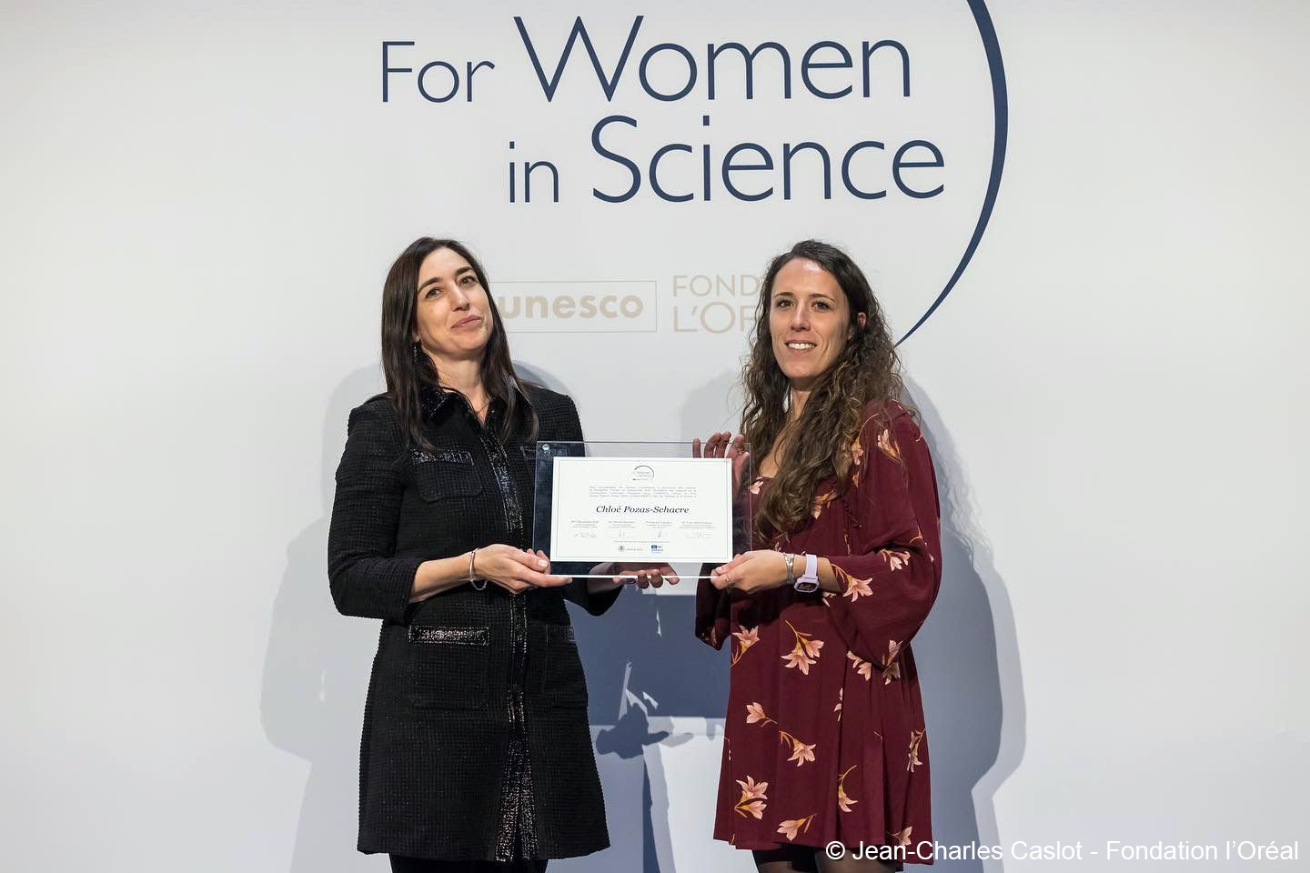
Chloé receiving her Fondation L’Oreal-UNESCO French Young Talents 2022 award.
Photo: Jean-Charles Caslot

Proliferation of Turbinaria ornata on Moorea’s fore reef.
Photo: Yannick Chancerelle
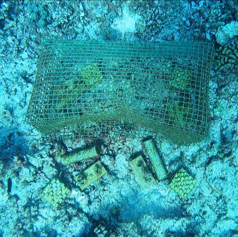
Portion of an experimental plot showing cage, nutrient diffusers, recruitment tiles and transplanted juvenile algae.
Photo: Manon Marco

Manola Bejarano checking out the MECATUR experiment.
Photo: Manon Marco
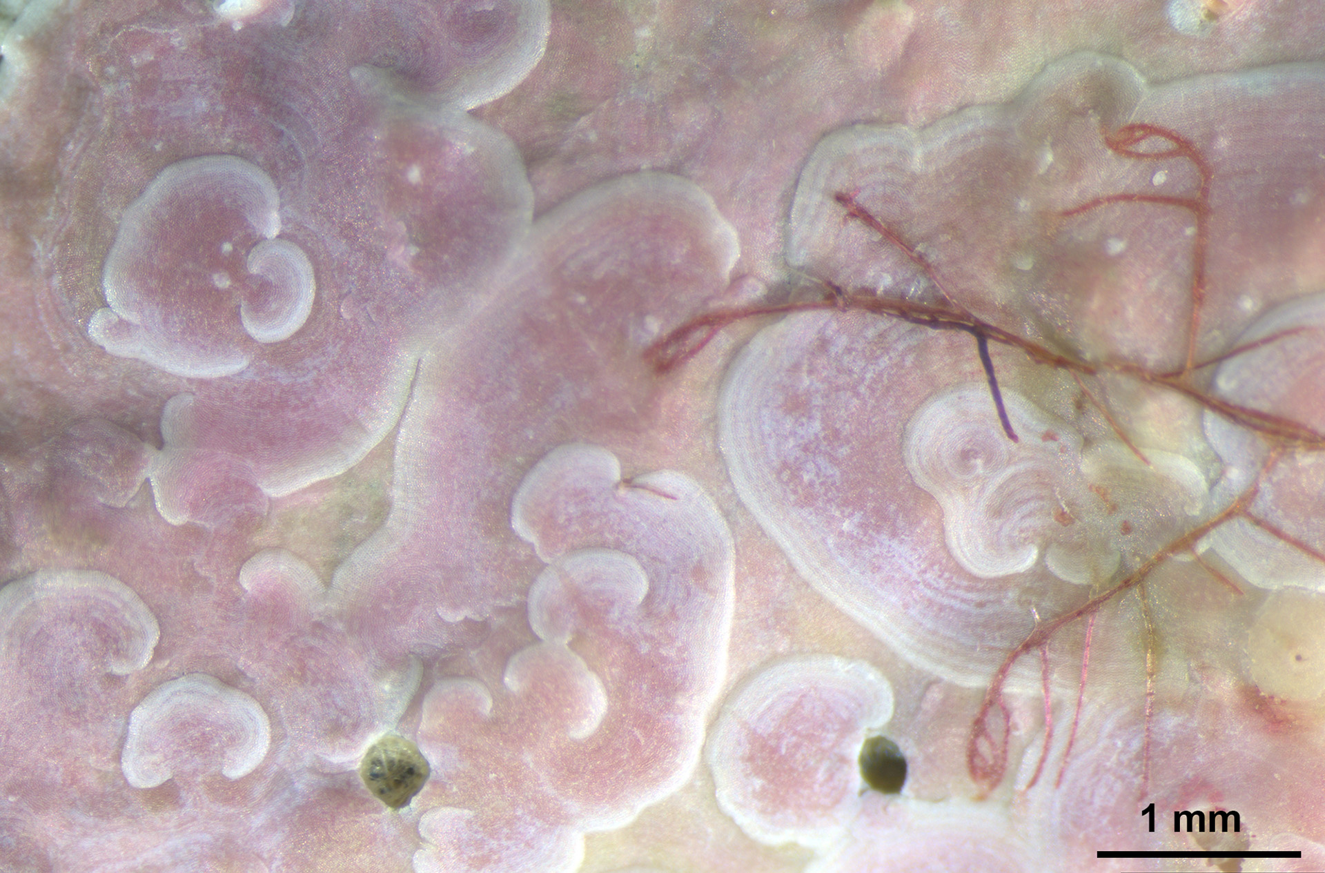
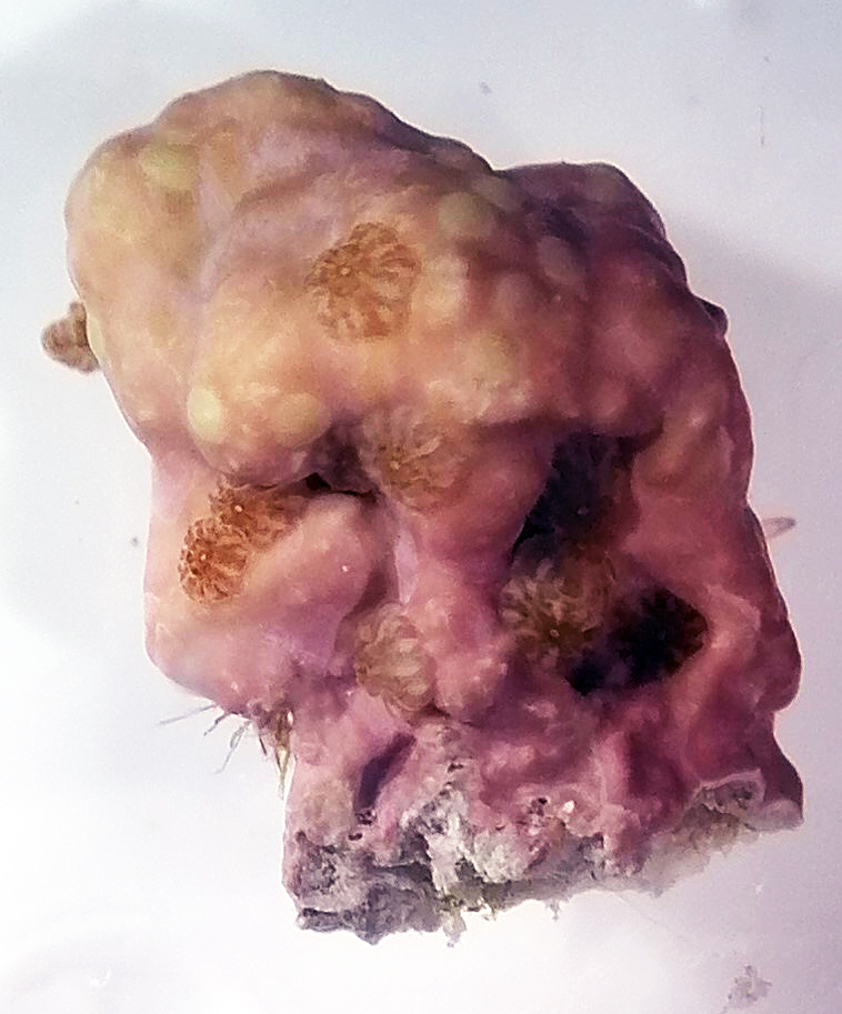
Lithophyllum sp. with swirling layers.
Photo: Maggy Nugues
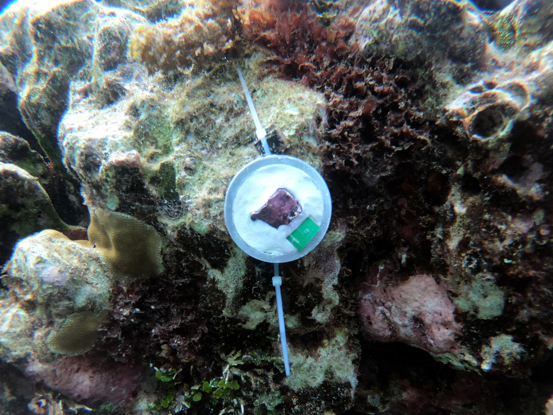
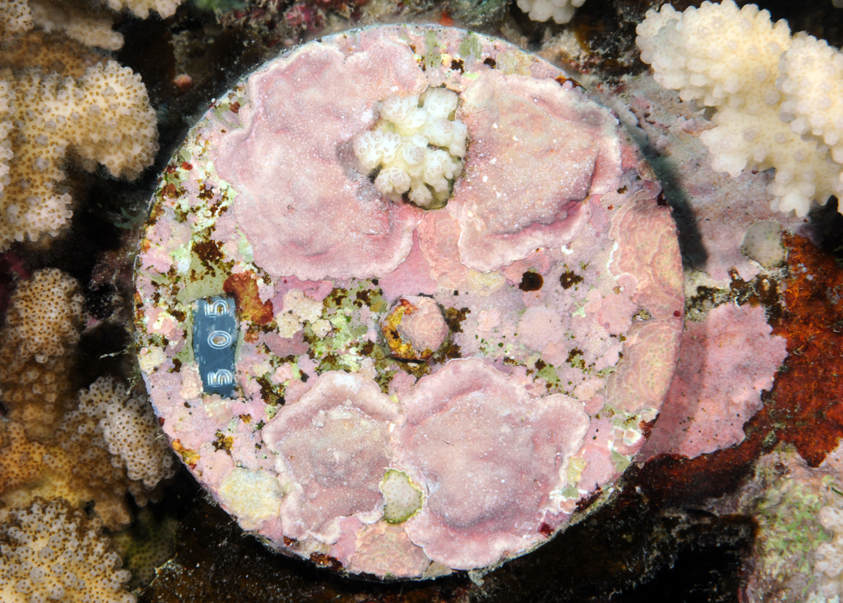
PVC rings with CCA chips used to estimate growth and calcification.
Photo: Camille Vizon

PVC rings with CCA chips used to estimate growth and calcification.
Photo: Camille Vizon
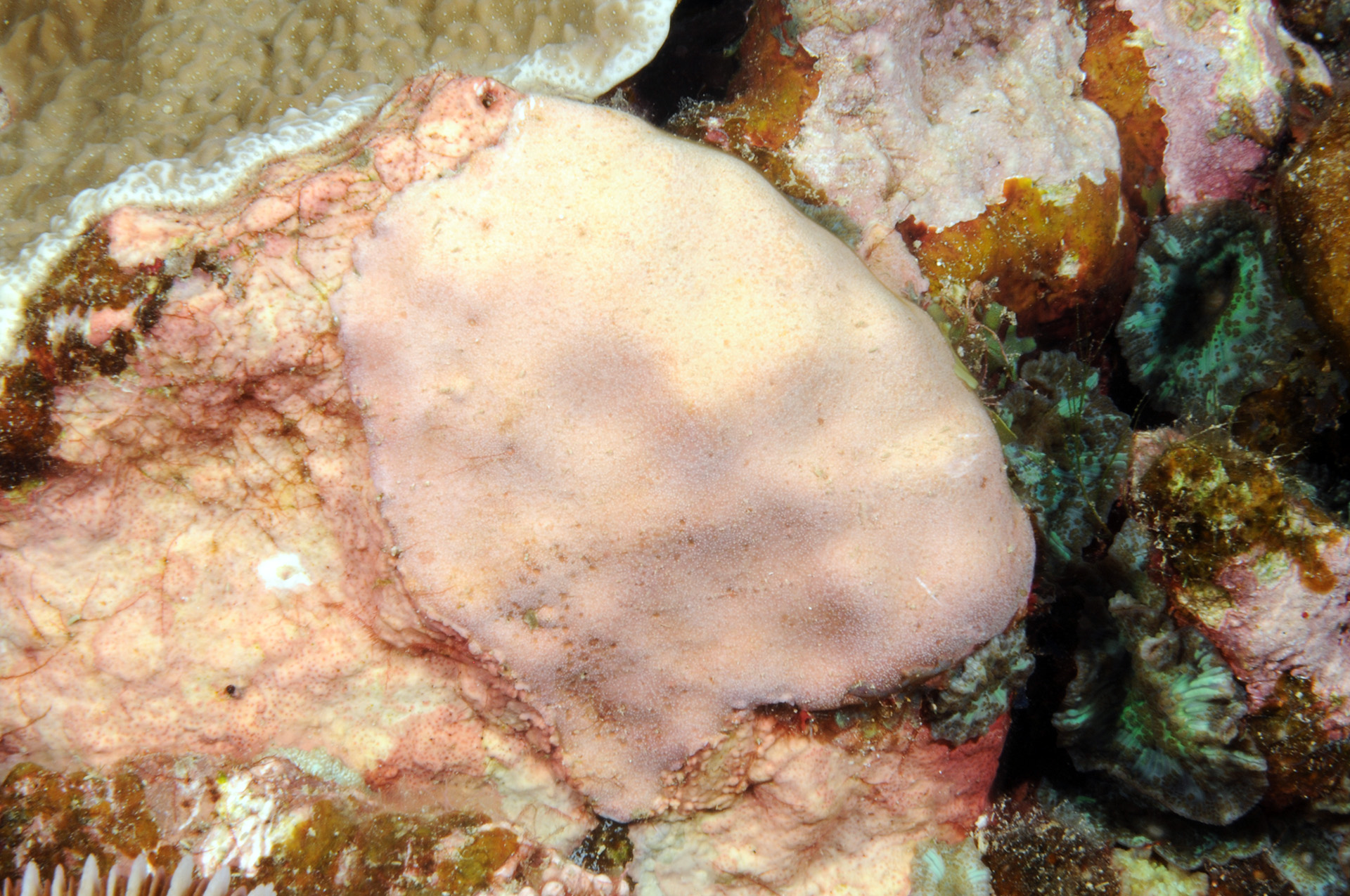
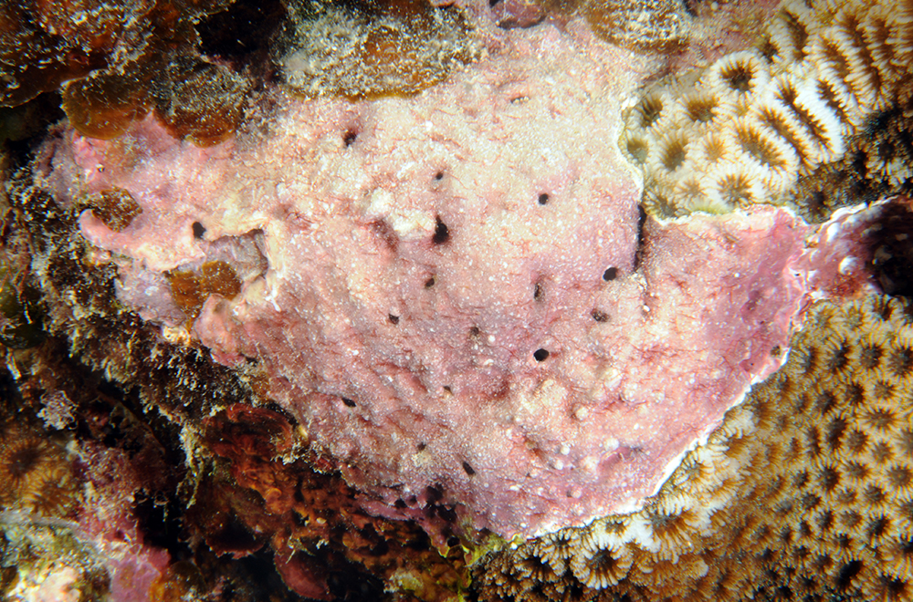
The crustose coralline algae Porolithon onkodes. Photo: Maggy Nugues
Hugo Bischoff removing macroalgae. Photo: Chloé Pozas-schacre
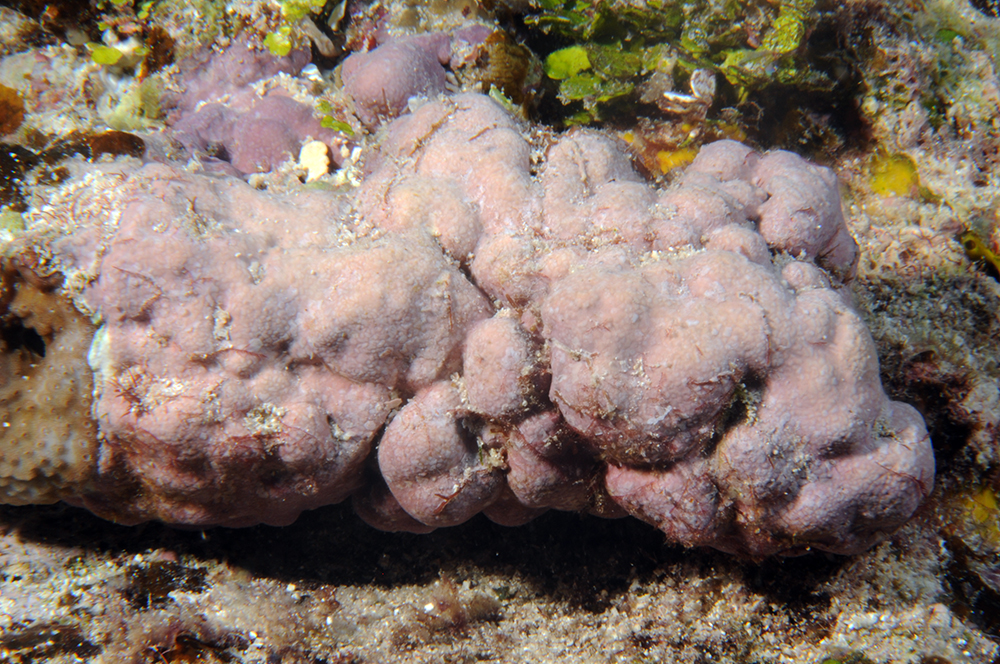
The crustose coralline alga Dawsoniolithon spp. Photo: Maggy Nugues
The crustose coralline alga Hydrolithon spp. Photo: Maggy Nugues
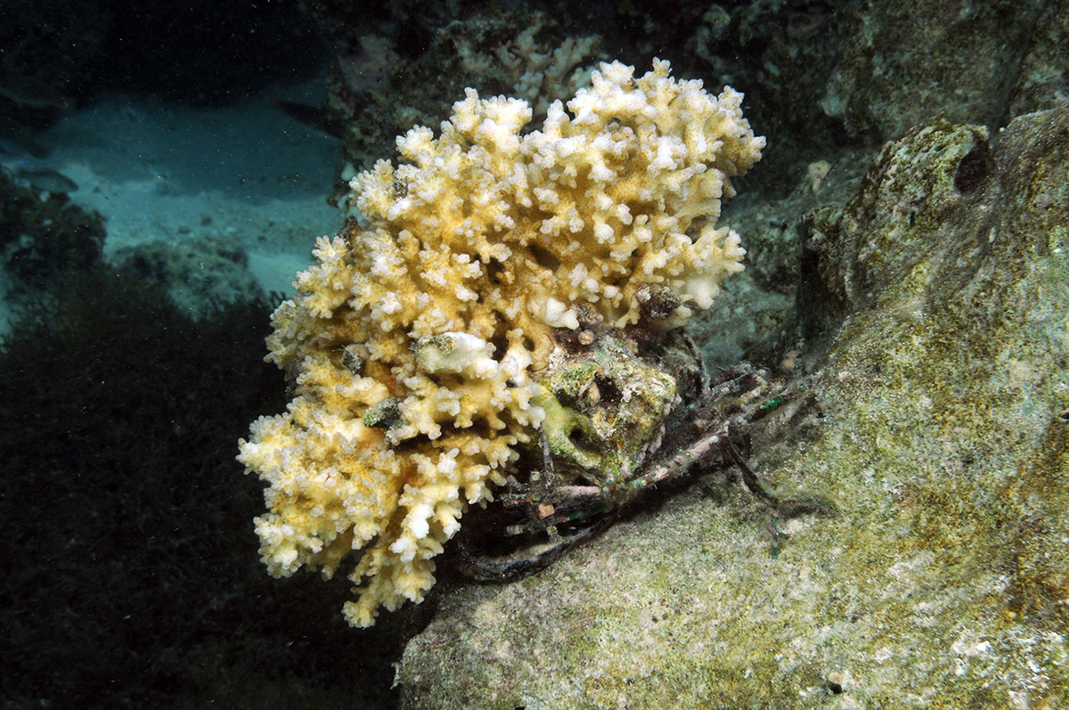
Pocillopora acuta colony. Photo: Maggy Nugues
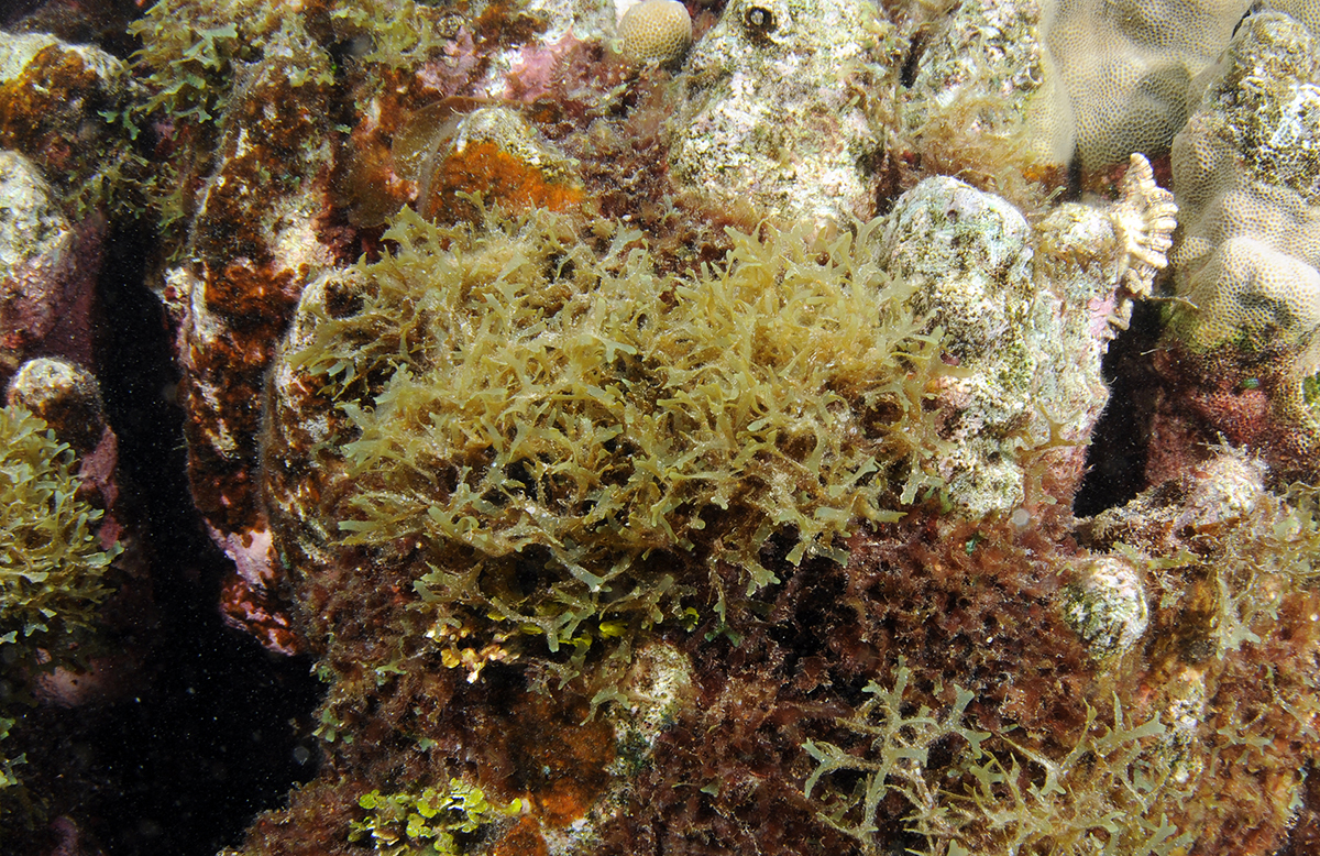
The Y branched alga Dictyota bartayresiana. Photo: Maggy Nugues
Funding source : ANR